Definition | Treatment | Management of Disease | Diagnosis | Symptoms | Etiology | Pathophysiology | Procedure


Management of Cough

The best management of cough is best to specific drug delivery to the etiology. Three forms of management of cough are:

1. Without the drug delivery
Cases with a cough without the interference caused by acute illness and heal itself usually does not need medication.
2. Specific Treatment
This treatment is given to the causes of cough.
If the cause of cough is known then the treatment should be directed towards the cause. With an integrated diagnostic evaluation, in almost all patients can be a known cause of chronic cough.
Specific treatment depends on the etiology or the cough mechanism. Asthma treated with bronchodilators or corticosteroids. Post nasal drip due to sinusitis treated with antibiotics, nasal spray and antihistamine-decongestant combinations, post nasal drip due to allergies or non allergic rhinitis dealt with avoiding environments that have the precipitating factors and antihistamine-decongestant combinations.
Gastroesophageal reflux treated by elevating the head, dietary modifications, antacids and cimetidine. Cough in chronic bronchitis treated by stopping smoking. Antibiotics are given to pneumonia, sarcoidosis treated with corticosteroids and cough in congestive heart failure with digoxin and furosemide.
Specific treatment also may include surgery such as pulmonary resection in lung cancer, polypectomi, remove hair from the outer ear canal.
Cases with a cough without the interference caused by acute illness and heal itself usually does not need medication.
3. Symptomatic treatment
Given both to patients who can not determined the cause of the cough as well as to patients who cough is a nuisance, not working properly and can potentially cause complications.
Symptomatic treatment is given if:
The cause of cough is certainly not known, so that specific treatment can not be given.
Coughing is not functioning properly and its complications endanger the patient.
Drugs used for symptomatic treatment of two types namely:
a. Antitussive
Antitussive is a medication that suppress the cough reflex, used in respiratory disorders and unproductive coughs due to irritated skin.
In general, based on place of work is divided into antitussive  drug that works in the peripheral and central antitussive who works at. Working in the central antitussive divided into non-narcotic and narcotic.
b. Mucokinesis
A pathologic fluid retention in the airway is called mucostasis. Drugs that are used to handle the situation called mucokinesis.

See more : 

Cough - Causes and Complications


Cough Reflex
Cough reflex consists of five main components, ie, cough receptors, afferent nerve fibers, central cough, nervous system and efferent effectors. A cough begins with a stimulus on cough receptor. These receptors are non-myelinated nerve fibers in the form of finely located both inside and outside the thoracic cavity. Located within the thoracic cavity, among others, contained in the larynx, trachea, bronchi and the pleura. Will decrease the number of receptors on the branches of a small bronchus, and a large number of receptors found in the larynx, trachea, carina and bronchus branching region. The receptors are also found even in the ear canal, stomach, hilum, paranasalis sinus, pericardial and diaphragm.
The most important afferents exist in the branch of the vagus, which excitatory flow of the larynx, trachea, bronchus, pleura, stomach, and also stimulate the ear through the Arnold branch of n. Vagus. Trigeminal nerve stimulation of the sinus paranasalis channeling, channeling glosofaringeus nerve stimulation of the pharynx and channel phrenic nerve stimulation of the pericardium and diaphragm.

Causes of Cough
Cough in outline can be caused by excitatory as follows:
Inflammatory stimuli such as mucosal edema with a lot of tracheobronchial secretions.
Mechanical stimuli such as foreign body in airway foreign body such as the respiratory tract, post nasal drip, retention of bronchopulmonary secretions.
Temperature stimuli such as cigarette smoke (an oxidant), heat / cold, gas inhalation.
Psychogenic stimuli.

Some causes of cough
  • Irritant
- Cigarettes
- Smoke
- SO2
- Gas in the workplace
  • Mechanical
- Retention of bronchopulmonary secretions
- Foreign body in respiratory tract
- Post nasal drip
- Aspiration
  • Obstructive Pulmonary Disease
- Chronic Bronchitis
- Asthma
- Emphysema
- Firbrosis cystic
- Bronchiectasis

  • Restrictive Lung Disease
- Pneumoconiosis
- Diseases of collagen
- Granulomatous Disease
  • Infection
- Acute Laryngitis
- Acute Brochitis
- Pneumonia
- Pleurisy
- Pericarditis

  • Tumor
- Laryngeal tumors
- Lung Tumors

  • Psychogenic
  • Other


Complications of Cough
At the time of coughing intrathoracic pressure rises to 300 mmHg. Pressure elevation is required to produce an effective cough, but this can lead to complications in the lungs, musculoskeletal, cardiovascular system and central nervous system.

Pneumomediastinum may arise in the lung, may also occur pneumoperitonium and pneumoretropritonium but this is very rare. Another complication was pneumothorax and emphysema, complications muskuloskletal, broken ribs, ruptured abdominal rectus muscle. Cardiovascular complications may include bradycardia, subconjungtiva vein laceration, nose and anus as well as cardiac arrest.

In the central nervous system can occur cough syncope, due to increased intrathoracic pressure occurs reflex vasodilation of systemic arteries and veins. This leads to decreased cardiac output and sometimes berkibat low arterial pressure resulting in loss of consciousness. Syncope occurred a few seconds after the paroxysmal cough.

Can also occur among other constitutional symptoms of insomnia, fatigue, decreased appetite, vomiting, elevated body temperature and headaches. Another complication is urinary incontinence, hernias and prolapse of the vagina.

Cough - Definition and Mechanisms

Cough
Cough is a symptom of the most common disease in which the prevalence was found in about 15% in children and 20% in adults. One out of ten patients who visited the doctor's office each year has a chief complaint of cough. Coughing can cause bad feelings, sleep disorders, affecting daily activities and reduce quality of life.

Cough is a complex physiological reflex that protects the lung from mechanical trauma, chemical and temperature. Coughing is also a natural defense mechanism of lung airway to keep it clean and open to the street:
Prevent the entry of foreign objects into the respiratory tract.
Remove foreign objects or abnormal secretions from the respiratory tract.
Coughing becomes physiologically when perceived as a nuisance. Such a cough is often a sign of a disease within or outside the lungs and sometimes are the early symptoms of a disease. Cough may be very significant on the transmission of disease through the air (air-borne infection). Cough is one of the symptoms of respiratory tract diseases in addition to tightness, wheezing, and chest pain. Often the cough is a problem faced by physicians in their daily work. The reason is very diverse and the introduction of the pathophysiology of cough would be very helpful in establishing the diagnosis and prevention of people with coughs. Research shows that in patients with acquired chronic cough 628 cough up to 761 times / day. Patients with pulmonary TB coughs number about 327 times / day and patients with influenza-even up to 154.4 times / day.
Epidemiological studies have shown that many chronic cough associated with smoking. Twenty-five percent of those who smoked 1 / 2 pack / day will have to cough, while the patients who smoked one pack per day will be found to be approximately 50% of chronic cough. Most of the heavy smokers who smoked 2 packs / day will complain of chronic cough. Large-scale research in the U.S. also found that 22% of non-smokers also suffer from cough, among others, caused by chronic diseases, air pollution and others. Coughing can also cause various complications such as pneumothorax, pneumomediastinum, headache, fainting, disc herniation, inguinal hernia, broken ribs, bleeding subkonjungtiva, and urinary incontinence.

Definition
Cough in Latin called tussis is a reflex that can occur suddenly and often repeated that aims to help clear mucus from the respiratory tract of, irritants, foreign particles and microbes. Coughing can happen voluntarily or involuntarily.
Coughing is a reflex action in the respiratory tract that is used to clean the upper airways. Chronic cough lasts more than 8 weeks are common in the community. Causes include smoking, exposure to cigarette smoke, and exposure to environmental pollutants, especially particulates.

Mechanism of Occurrence of Cough
Cough starts from a cough receptor stimulation. These receptors are non-myelinated nerve fibers in the form of finely located both inside and outside the thoracic cavity. Located within the thoracic cavity, among others, contained in the larynx, trachea, bronchus, and pleura. Will decrease the number of receptors on the branches of a small bronchus, and a large number of receptors in the can in the larynx, trachea, carina and bronchus branching region. The receptors are also found even in the ear canal, stomach, hilum, paranasalis sinus, pericardial, and diaphragm.
Mechanism Of Cough
Afferent fibers are the most important branch of the vagus on the drain stimuli from larynx, trachea, bronchus, pleura, stomach, and also stimulation of the ear through the Arnold branch of the vagus. Trigeminal nerve stimulation of the sinus paranasalis channel, glosofaringeus nerve, excitatory channel of the pharynx and channel phrenic nerve stimulation of the pericardium and diaphragm.

By excitatory afferent fibers was taken to the cough center located in the medulla, near the center of the respiratory and vomiting centers. Then from here by afferent fibers of the vagus, phrenic nerve, intercostal and lumbar nerves, the trigeminal nerve, facial nerve, nerve hipoglosus, and others headed to the effector. These effector standing of the muscles of the larynx, trachea, bronchi, diaphragm, intercostal muscles, and others. In the area of ​​this effector mechanism of cough ensued.


Phase of Cough
Basically the mechanism of cough can be divided into four phases, namely:


1. Phase irritation
Irritation of one sensory vagus nerve in the larynx, trachea, large bronchi, or afferent fibers from the pharyngeal branch glosofaringeus nerve can cause coughing. Coughing also arise when the cough receptors in the lining of the pharynx and esophagus, pleural cavity and external ear canal is stimulated.

2. Phase of inspiration
In the inspiration phase of the glottis is wide open due to reflex muscle contraction abduktor aritenoidea cartilage. Inspiration occurs in and quickly, so the air quickly and in large quantities into the lungs. This is accompanied terfiksirnya rib due to muscle contraction under the thorax, abdomen and diaphragm, so that the lateral dimension enlarged breasts lead to increased lung volume. The entry of air into the lungs by the number of lots of benefits that will strengthen the expiratory phase resulting in faster and stronger and reduce the air cavity is closed so as to produce a potential cleaning mechanism.

3. Compression phase
This phase begins with the closing of the glottis due to contraction of the adductor muscle aritenoidea cartilage, glottis closed for 0.2 seconds. In this phase the intrathoracic pressure rises to 300 cmH2O to place an effective cough. Pleural pressure remained elevated for 0.5 seconds after the glottis open. Coughing can occur without glottis closure because of expiratory muscles to increase intrathoracic pressure although the glottis remains open.

4. Expiratory phase / expulsion
In this phase the glottis opens suddenly due to active contraction of expiratory muscles, so there was spending large amounts of air at high speed accompanied by spending foreign objects and other materials. Movement glottis, respiratory muscles and the branches of the bronchus is important in the mechanism of cough phase and this phase of cough is actually happening. Cough sound varies greatly due to vibration secretions in the respiratory tract or the vibration of vocal cords

Cough - Causes and Complications
Management of Cough

Lung Defense Mechanism


Lung defense mechanisms are very important in explaining the occurrence of respiratory infections. lung has defense mechanisms to prevent bacteria from entering into the lungs. cleaning mechanism are:
1. Cleaning mechanism in the conductive airways, including:
• Reepitelisasi airway
• The flow of mucus on the surface epithelium
• Bacteria naturally or "epithelial-cell binding site analogue"
• Local humoral factors (IgG and IgA)
• Components of the local microbial
• Mucosilliar transport system
• Sneezing and coughing reflexes

Upper respiratory tract (nasopharynx and oropharynx) is a defense mechanism through the barrier against the entry of the anatomy and mechanism of pathogenic microorganisms. Cilia and mucus push microorganisms out of the way coughed or swallowed.
If there is dysfunction of cilia as in Kartagener's syndrome, the use of nasogastric tube and pipe nasotracheal long can disrupt the flow of secretions that have been contaminated with pathogenic bacteria. In these circumstances may occur nosocomial infection or "Hospital Acquired Pneumonia".

2. Cleaning mechanism in "Respiratory airway exchange", include:
• The fluid lining the alveolar surfactant include
• The system of local humoral immunity (IgG)
• Alveolar Macrophages and inflammatory mediators
• Withdrawal of neutrophils
Humoral immune system plays an important role in lung defense mechanisms (upper airway). IgA is one part of the nasal secretions (10% of the total protein nasal secretions). Patients with IgA deficiency have an increased risk for upper respiratory tract infection recurring yan. Colonization of bacteria that often hold the upper airway often removing and damaging IgA proteolytic enzymes. Gram-negative bacteria (Pseudomonas aeruginosa, E.colli, Serratia spp, Proteus spp, and Klebsiella pneumoniae) has the ability to destroy IgA. Deficiencies and damage to any component of upper airway defense led to the colonization of pathogenic bacteria as facilities for lower respiratory tract infection.

3. Cleaning mechanism in the subglottic airways
Subglottic airway defense mechanisms composed of anatomic, mechanical, humoral and cellular components. Mechanism of closure of the glottis and cough reflex is the main defense against the aspirate from the oropharynx. In case of malfunctioning of the glottis it dangerous for lower respiratory tract that are normally sterile. Nasogastric tube mounting action, tool tracheostomy facilitate the entry of bacterial pathogens directly into the lower respiratory tract. Impaired mucociliary function may facilitate the entry of pathogenic bacteria into the lower respiratory tract, even acute infection by Mycoplasma pneumoniae, Haemophilus influenzae and viruses can damage the cilia movement.

4. Cleaning mechanism in the "respiratory airway gas exchange"
Bronchioles and alveol have a defense mechanism as follows:
• The liquid that coats alveol:
a. Surfactants
A fat-rich glycoprotein, consists of several components of SP-A, SP-B, SP-C, SP-D that serves to strengthen phagocytosis and killing of bacteria by macrophages.
b. Anti-bacterial Activity (non specific) : FFA, lysozyme, iron binding protein.
• IgG (IgG1 and IgG2 subset that serves as an opsonin)
• alveolar macrophages that act as the first defense mechanism
• Serves to attract PMN leucocytes into the alveoli (there GNB infections, Pseudomonas aeruginosa)
• Mediator biology
Ability to attract PMN to the airway, including C5a, the production of alveolar macrophages, cytokines, leucotrienes
Tags : Defense Mechanism of lung, Defense Mechanisms of the Respiratory SystemPulmonary defence mechanisms

Anesthesia Patients With Asthma

Pre-operation / Pre-anesthesia Assessment
Anamnesis
History about whether patients had received previous anesthesia is very important to know if there are things that need special attention, for example: allergies, nausea, vomiting, itching or shortness of breath after the surgery, so that we can design the next anesthetic well.
Physical examination
State of dentition examination, action open mouth, the tongue is relatively large is very important to know whether the action would make it difficult laryngoscopy intubation. Another routine systemically check about the general course should not be missed such as inspection, palpation, percussion and auscultation of all organ systems of patients. Examination of inspection, palpation, percussion and auscultation on the cardiopulmonary system is a major clinical examination of much help in the assessment of asthma. With this examination can be known frequency of breathing, respiratory pattern, presence of wheezing / ronchi.
ECG examination
In addition to knowing about the state / heart disease, especially the picture ECG can also determine the influence of lung function.
Radiological examination
Includes images of the thorax and CT-scan (if necessary). This examination is not to assess lung function disorders but it is important for supporting the diagnosis of pulmonary disease, signs of lung hyperinflation and congestive heart disease, also to help determine abnormalities in the mediastinal cavity (CT-scan).
Laboratory examination
Include routine blood tests, blood gas analysis of blood sugar.
Pulmonary Physiology Tests
  • Without tools: although simple but can provide information on respiratory function and are useful as an assessment of "fronss for operation" such as the ability to climb stairs without shortness of breath while talking. Snider match test: the ability to hold breath for 30 seconds.
  • Using a spirometer.

Physical Status Classification
Classification is commonly used to assess a person's physical fitness is derived from The American Society of Anaesthesiologist (ASA). Physical classification is not a forecasting tool ratio of anesthesia, because the side effects of anesthesia can not be separated from the side effects of surgery.
  • Class I   : Patients healthy organic, physiological, psychiatric, biochemistry.
  • Class II  : Patients with mild or moderate systemic disease.
  • Class III : Patients with severe systemic disease, so that routine activities are limited.
  • Class IV : Patients with severe systemic disease can not perform routine activities and the disease is life threatening at any moment.
  • Class V  : dying patient who is expected with or without surgery her life would not be more than 24 hours
  • In Cito or emergency surgery is usually imprinted letter E.

Pre-Operative Preparation
Fasting
Laryngeal reflexes decreased during anesthesia. Regurgitation of gastric contents and impurities contained in the airway is a major risk in patients undergoing anesthesia. To minimize this risk, all patients scheduled for elective surgery should dipantangkan of oral input (fasting) during a certain period before induction of anesthesia.

Perioperative Management
For anesthesia and elective surgery in patients with a history of asthma, then asthma should be controlled and the patient was not suffering from an infection or severe wheezing attacks. If a patient takes medication on a regular basis, then the drug should not be stopped. Particular attention should be given pad of patients who use steroids, systemically or by inhaler.
Bronchospasme may be induced by anxiety, pain, drugs, endotracheal intubation, foreign body or irritation. Drugs that are contraindicated are: tubocurarrine and anticolinesterase, Sodium thiopental (Pentothal™), morphine, papaverin, trimethaphan and beta blockers.
Many drugs that can be used as a premedication such as diazepam, pethidine, promethazine and atropine, an estimated free from bronchospatic activity. Bronchodilator and steroid therapy is continued.

Anesthesia Techniques
Whenever possible, choose regional anesthesia with continuous epidural block with a low of 1% lidocaine (analgesia only) so that the respiratory muscles are not disrupted.
If general anesthesia is required then given premedication with antihistamines such as promethazine together with hydrocortisone 100 mg. What is important to avoid laryngoscopy and intubation with a shallow anesthesia, because it can cause bronchospasm. Ketamine is good enough for intravenous induction, because it is a bronchodilator. For a brief action, you should use a face mask after induction techniques and avoid intubation. Use of oxygen with a concentration of 30% or more for air inspiration. If intubation is required, then deepened with inhalation anesthesia, and then do the intubation without muscle relaxants. In the anesthetized patient in laryngoscopy can be done without causing bronchospasm when intubated. Vecuronium may be administered as a muscle relaxant is good because it does not release histamine. Ether and halothane is bronchodilator good, but ether has the advantage, that is if there is bronchospasm, epinephrine (0.5 mg subcutaneously) can be administered safely (but this is dangerous if given in conjunction with halothane or trichlorethylene, because it can cause heart rhythm disturbances due to effects of catecholamines). As an alternative to epinephrine, aminophylline 250 mg can be administered intravenously slowly to mature; drug is compatible with all inhaled medication.
At the end of the action when using intubation, extubation done on his side and with anesthesia in, because stimulation of the larynx can cause bronchospasm.

Post Operative Care
Provision of adequate analgesia postoperative care is vital. Adequate oxygenation. Maintenance intravenous fluids. Usually anti-asthma drugs are still needed. Form of steroid drugs given intravenously as a temporary substitute for oral medications and inhalers brochodilator nebulizer as a substitute if the patient can not breathe in, or not yet maximal lung function after surgery. In the event of failure of achieving adequate ventilation and oxygenation after surgery, the patient to go to Intensive Care Unit (ICU).

Tags : Anesthesia and Asthma, Anesthesia With Asthma, Ketamine and asthma,

Pulmonary Edema

Definitions
pulmonary edema definition, pulmonary oedema, flash pulmonary edema, acute pulmonary edema, pulmonary edema symptoms, pulmonary edema causes, pulmonary edema treatment, lung edema, altitude pulmonary edema,
Edema, in general, means swelling. This typically occurs when fluid from the inside of blood vessels leak out of blood vessels into surrounding tissues, causing swelling. This can occur because of too much pressure in blood vessels or there is not enough proteins in the blood stream to hold the liquid in the plasma (part of blood that does not contain any blood cells).
Pulmonary Edema is the term used when edema occurs in the lungs. The area immediately outside of the small blood vessels in the lung occupied by air pockets are very small, called alveoli. This is where oxygen from the air through which blood is taken by, and carbon dioxide in the blood released into the alveoli to exhaled out. Normal alveoli have very thin walls that allow air exchange, and liquids are usually kept away from the walls of the alveoli-dindig unless it loses its integrity.
Pulmonary edema is an accumulation of fluid in the lungs due to sudden increase in intravascular pressure. Pulmonary edema is due to the flow of fluid from the blood into the space next to interstisial pulmonary alveoli of the lungs, exceeding the liquid back into the blood stream or through lymphatic channels.
Pulmonary edema is a condition caused by excess fluid in the lungs. This fluid collects in the air sacs in the lungs of many, making it difficult to breathe. In most cases, heart problems cause pulmonary edema. But fluid can accumulate due to other reasons, including pneumonia, exposure to certain toxins and drugs, and sports or live at high altitude.
Pulmonary edema is the term used when edema occurs in the lungs. Pulmonary edema is a condition caused by excess fluid in the lung.

Causes
1. Imbalance of Starling Forces:
a) Increased pulmonary capillary pressure:
  • Increased pulmonary venous pressure in the absence of left ventricular dysfunction (mitral stenosis).
  • Increased pulmonary venous pressure secondary because of impaired left ventricular function.
  • Increased pulmonary capillary pressure secondary because of increased pulmonary artery pressure (over-perfusion pulmonary edema).
b) Decrease in plasma oncotic pressure.
  • Hypoalbuminemia secondary because of kidney disease, liver, protein-losing enteropaday, dermatological diseases or nutritional diseases.
c) Increased negative pressure intersisial:
  • Taking too fast pneumothorax or pleural effusion (unilateral).
  • A highly negative pleural pressure due to acute airway obstruction in conjunction with an increase in end-expiratory volume (asthma).
d) Increased oncotic pressure intersisial.

  • Until now there has been no example of a trial or clinic.
2. Changes in alveolar-capillary membrane permeability (Adult Respiratory Distress Syndrome)

a) Pneumonia (bacteria, viruses, parasites).
b) The material inhaled toxic (phosgene, ozone, chlorine, smoke, Teflon ®, NO2, etc.).
c) Foreign material in the circulation (snake venom, bacterial endotoxins, alloxan, alpha-naphthyl Thiourea).
d) Aspiration of gastric acid.
e) Acute radiation pneumonitis.
f) Material endogenous vasoactive (histamine, kinin).
g) Disseminated intravascular coagulation.
h) Immunology: hypersensitivity pneumonitis, drug nitrofurantoin, leukoagglutinin.
i) Shock Lung therefore outside the thoracic trauma.
j) Bleeding Acute Pancreatitis.

3. Lymphatic insufficiency:
a) Post Lung Transplant.
b) Lymphangitic carcinomatosis.
c) fibrosing lymphangitis (silicosis).
4. Not known / not clear
a) High Altitude Pulmonary Edema.
b) Neurogenic Pulmonary Edema.
c) Narcotic overdose.
d) Pulmonary embolism.
e) Eclampsia
f) Post cardioversion.
g) Post Anesthesia.
h) Post Cardiopulmonary Bypass.
Classification
Based on the cause, is divided into 2 pulmonary edema, cardiogenic and non-cardiogenic. It is important to know because its treatment is very different. Cardiogenic Pulmonary Edema due to Left Heart Terrible any reason. Acute Cardiogenic Pulmonary Edema is caused by the presence of Acute Left Heart Sucks. But with the precipitation factors, may occur also in patients with Chronic Left Heart Sucks.

» Cardiogenic pulmonary edema
Edema, cardiogenic pulmonary edema is caused by abnormalities in the heart organ. For example, the heart does not work properly as the heart pumps are not good or strong the heart is not pumping anymore.
Cardiogenic pulmonary edema resulting from high pressure in blood vessels of the lungs caused by poor heart function. Congestive heart failure caused by poor cardiac pump function (coming from a variety of causes such as arrhythmias and diseases or weakness of the heart muscle), heart attacks, or heart valves can lead to abnormal accumulation of more than the usual amount of blood in blood vessels of the lungs. This can, in turn, causes the fluid from the blood vessels are pushed out into the alveoli when the pressure is growing.

» Non-cardiogenic pulmonary edema
Non-cardiogenic pulmonary edema is that edema is usually caused by the following:

  • Acute respiratory distress syndrome (ARDS). In ARDS, the integrity of the alveoli become compromised as a result of an underlying inflammatory response, and this menurus on a leaky alveoli that can be filled with fluid from blood vessels.
  • A potentially serious condition caused by severe infections, trauma, lung injury, inhalation of toxins, lung infections, smoking cocaine, or radiation to the lungs.
  • Renal failure and inability to remove fluid from the body can cause a buildup of fluid in blood vessels, resulting in pulmonary edema. In people with advanced kidney failure, dialysis may be necessary to remove excess body fluids.
  • High altitude pulmonary edema, which can occur due to a rapid rise to high altitudes over 10,000 feet.
  • Traumatic brain, bleeding in the brain (intracranial hemorrhage), severe seizures, or brain surgery can sometimes result in accumulation of fluid in the lungs, causing neurogenic pulmonary edema.
  • Lung expanding rapidly can sometimes lead to a re-expansion pulmonary edema. This may occur in cases when a collapsed lung (pneumothorax) or large amounts of fluid around the lungs (pleural effusion) was issued, resulting in rapid expansion of the lung. This can result in pulmonary edema only on the affected side (unilateral pulmonary edema).
  • Rarely, overdose on heroin or methadone can lead to pulmonary edema. An overdose of aspirin or the use of high doses of aspirin can lead to chronic aspirin intoxication, especially in the elderly, which may cause pulmonary edema.
  • Other causes are less frequent than non-cardiogenic pulmonary edema may include pulmonary embolism (blood clot that had walked into the lungs), acute lung injury related to transfusion or transfusion-related acute lung injury (TRALI), some infections -viral infection, or eclampsia in pregnant women.

Pathophysiology
Pulmonary edema occurs when the alveoli are filled with excess fluid that seeps out of blood vessels in the lungs instead of air. This can cause problems with gas exchange (oxygen and carbon dioxide), resulting in difficulty breathing and poor blood pengoksigenan. Occasionally, this can be referred to as "water in the lungs" when describing this condition in patients. Pulmonary edema can be caused by many different factors. He can be connected in heart failure, called cardiogenic pulmonary edema, or linked to other causes, referred to as non-cardiogenic pulmonary edema.

Clinical Manifestations
The most common symptoms of pulmonary edema is shortness of breath. This is probably a gradual onset if the process develops slowly, or he may have a sudden onset in cases of acute pulmonary edema. Other common symptoms may include fatigue, shortness of breath develop faster than normal with usual activities (Dyspnea on Exertion), rapid breathing (tachypnea), dizziness, or weakness.
Low blood oxygen levels (hypoxia) may be detected in patients with pulmonary edema. Furthermore, upon examination of the lungs with a stethoscope, the doctor may hear abnormal lung sounds, rales or crackles crate (short boiling sounds disjointed, which corresponds to the muncratan fluid in the alveoli during breathing).
Clinical manifestations Pulmonary Edema specifically also divided into 3 stages:
Stage 1.
The existence of small blood vessels distended and prominent lung would improve gas exchange in lungs and slightly increase the capacity of diffusion of CO gas. Complaints at this stage may just be a shortness of breath while working. Physical examination is also not clearly found abnormalities, except maybe the inspiration for ronkhi at the time of opening of the closed airway during inspiration.
Stage 2.
At this stage intersisial pulmonary edema. Limit pulmonary blood vessels become blurred, so too the hilum also become blurred and interlobularis thickened septa (Kerley B lines). By accumulation of fluid in the loose network of inter-sisial, will further reduce the small airways, especially in the basal area because of the influence of gravity. Might also occur bronkhokonstriksi reflex. Often there is takhipnea. Although this is a sign of impaired left ventricular function, but takhipnea also help pump lymph flow is slowed so that the buildup of fluid intersisial. On examination there was little change in spirometry alone.
Stage 3.
At this stage of alveolar edema. Severely impaired gas exchange, hypoxemia and hypocapnia occur. Patients seem very crowded with reddish frothy cough. Vital capacity and other lung volume decreased markedly. Occurs right-to-left intrapulmonary shunt. Patients usually suffer from hypocapnia, but in severe cases can occur hypercapnia and acute respiratory acidemia. In this situation morphine has to be used with caution (Ingram and Braunwald, 1988).
Pam edema that occurs after acute myocardial infarction is usually due to capillary pulmonary hypertension. However, experiments on dogs that do arteriakoronaria ligation, pulmonary edema occurred despite normal pulmonary capillary wedge pressure, which can be prevented with indomethacin administration before. It is estimated that by inhibiting cyclic nucleotide phosphodiesterase or cyclooxygenase to reduce edema 'secondary to increased pulmonary alveolar-capillary permeability; on human beings still requires further research. Sometimes patients with acute myocardial infarction and pulmonary edema, capillary wedge pressure normal lungs; this is probably due to the slow clearance of edema fluid radiographically although pulmonary capillary pressure has gone down or alternatively in some patients an increase in alveolar-capillary permeability secondary lung because of the contents sekuncup low as in cardiogenic shock lung.

Diagnosis Supports
»Physical Examination

  • Central cyanosis. Shortness of breath by breath sounds like a frothy mucus.
  • Ronchi loud in the basal lung wet and then took up almost the entire lung field, sometimes accompanied by dry and expiratory Ronchi which extends a result of bronchospasm so-called cardiac asthma.
  • Tachycardia with S3 gallops.
  • Murmur when valve abnormalities.

»Electrocardiography. Can sinus tachycardia with left atrial hypertrophy or atrial fibrillation, depending on the cause of heart failure. Preview infarction, left ventricular hypertrophy or arrhythmias can be found.
»Laboratory

  • Low pO2 blood gas analysis, pCO2 initially low and then hypercapnia.
  • Enzymes cardiospesific increase if the cause of myocardial infarction.
  • Blood routine, urea, creatinine, electrolytes, urinalysis, thoracic images, ECG, cardiac enzymes (CK-MB, Troponin T), coronary angiography.

Photo thoracic Pulmonary edema is typically diagnosed by chest X-ray. Radiograph (X-ray) a normal chest consists of a centralized area that offends white heart and major blood vessels plus the bones of the vertebral column, with the lung fields showed as areas darker on each side, which surrounded by the bone structures of the chest wall.
X-ray chest with a typical pulmonary edema may show more white Tampakan in both lung fields than usual. The cases are more severe than pulmonary edema may show opacification (bleaching) is significant in the lung with minimal visualization of the lung fields are normal. Bleaching represents filling of the alveoli as a result of pulmonary edema, but it may provide minimal information about the possible underlying causes.
»Overview Radiology found:

  • Widening or thickening of the hilum (hilar vascular dilatation)
  • Increased lung pattern (more than 1 / 3 lateral)
  • Vascular cranialization
  • Bleak hilum (the limit is not clear)
  • Interstitial fibrosis (descriptions such as granulomas, small granulomas or miliary nodules)
»Preview the cause of heart failure echocardiography: valve abnormalities, ventricular hypertrophy (hypertension), Segmental wall motion abnormally (CHD), and generally found left ventricular dilatation and left atrium.

»Measurement of plasma B-type natriuretic peptide (BNP)
Other diagnostic tools used in assessing the underlying cause of pulmonary edema include the measurement of plasma B-type natriuretic peptide (BNP) or N-terminal pro-BNP. This is the marker proteins (hormones) that will arise in the blood caused by the stretching of the heart chambers. The increase of BNP nanograms (a billionth of a gram) per liter larger than a few hundred (300 or more) is very high suggesting cardiac pulmonary edema. On the other hand, the values ​​are less than 100 are essentially rule out heart failure as the cause.
»Pulmonary artery catheter (Swan-Ganz)
Pulmonary artery catheter (Swan-Ganz) is a long, thin tube (catheter) is inserted into large veins of the chest or neck and advanced through the room - right side of the heart chamber and placed into the pulmonary capillaries or pulmonary capillaries (branch -small branches of blood vessels of the lungs). This tool has the ability to directly measure the pressure in the pulmonary vessels, called pulmonary artery wedge pressure. Wedge pressure of 18 mmHg or higher is consistent with cardiogenic pulmonary edema, while wedge pressure of less than 18 mmHg usually support non-cardiogenic cause of pulmonary edema. Swan-Ganz catheter placement and interpretation of data is done only at the intensive care unit (ICU).

Differential Diagnosis 


Management
  • Semi-Fowler position.
  • Oxygen (40-50%) to 8 liters / minute if necessary with a mask.
  • If deteriorated (patients increasingly congested, takipneu, Ronchi added, can not be maintained PaO2 ≥ 60 mmHg with O2 concentration and high flow, CO2 retention, hypoventilation, or unable to adequately reduce the edema fluid), then performed endotracheal intubation, suctioning and ventilators.
  • Infusion emergency. Monitor blood pressure, ECG monitor, pulse oximetry if available.
  • Sublingual or intravenous nitroglycerin. Peroral nitroglycerin 0.4 to 0.6 mg every 50-10 minutes. If the systolic blood pressure> 95 mmHg may be given intravenous nitroglycerin starts dose of 3-5 ug / kg.
  • If it does not give satisfactory results it can be given IV Nitroprusid starting dose of 0.1 ug / kg / minute if they do not respond to nitrate, the dose is increased to obtain clinical improvement or until systolic blood pressure of 85-90 mmHg in patients who had had blood pressure normal or can be maintained as long as adequate perfusion to vital organs.
  • Morphine sulfate 3-5 mg iv, may be repeated every 25 minutes, a total dose of 15 mg (best avoided).
  • Diuretics Furosemide 40-80 mg IV bolus dose may be repeated or increased every 4 hours or continued drip continue to achieve urine output of 1 ml / kg / hour.
  • If necessary (blood pressure drop / mark hypoperfusion): Dopamine 2-5 ug / kg / min or dobutamine 2-10 ug / kg / min to stabilize hemodynamics. The dose may be increased according to clinical response or both.
  • Thrombolytic or revascularization in patients with myocardial infarction.
  • Ventilator in patients with severe hypoxia, acidosis / does not work with oxygen.
  • Operations on the complications of acute myocardial infarction, such as regurgitation, VSD, and ventricular wall rupture / corda tendinae.

Tags : pulmonary edema definition, pulmonary oedema, flash pulmonary edema, acute pulmonary edema, pulmonary edema symptoms, pulmonary edema causes, pulmonary edema treatment, lung edema, altitude pulmonary edema,

Respiratory Failure

Respiratory failure is the inability of the respiratory system to maintain normal blood oxygenation (PaO2), elimination of carbon dioxide (PaCO2) and pH Adequate ventilation problem caused by diffusion or perfusion.
Respiratory failure is inadequate gas exchange to hypoxia, hypercapnia (increased arterial carbon dioxide concentration), and acidosis.
Respiratory failure occurs when the exchange of O2 to CO2 in the lungs can not maintain the rate of O2 consumption and CO2 formation in the body's cells causing pO2 <> 2> 45 mmHg (hypercapnia) (Smeltzer, C Susane, 2001)


Causes / Etiology
1. The central nervous system depression
Resulting in respiratory failure due to inadequate ventilation. Respiratory centers that control breathing, located below the brain stem (pons and medulla) so slow and shallow breathing.
2. Primary neurological disorder
May hamper respiratory function. Impulses arising in the respiratory center spreads through the nerves that extend from the brainstem to the spinal nerves continue to receptors on the respiratory muscles. Diseases of the nervous disorders such as spinal cord, the muscles of respiration or neuromuslular meeting that occurs on breathing will greatly affect ventilation.
3. Pleural effusion, Pneumothorax and Hemothorax
It is a condition that interferes with ventilation through the inhibition of lung expansion. This condition is usually caused by defect underlying lung, pleural disease or trauma and injury and can cause respiratory failure.
4. Trauma
Caused by motor vehicles can be a cause of respiratory failure. Accidents resulting in head injuries, unconscious and bleeding from the nose and mouth can lead to upper airway obstruction and respiratory depression. Hemothorax, Pneumothorax and rib fractures may occur and possibly causing respiratory failure. Flail chest may occur and can lead to respiratory failure. The treatment is to correct the underlying pathology.
5. Acute pulmonary disease
Pneumonia caused by bacteria and viruses. Bronchial asthma, Atelectasis,Pulmonary embolism and pulmonary edema are some other conditions that cause respiratory failure.

Signs and Symptoms
Sign
a. Failed total breath
  • The flow of air in the mouth, the nose is not heard / felt
  • In spontaneous breathing movements supra visible retraction between the ribs and clavicles and no development of the chest on inspiration
b. Partial failure of breath
  • The sound of gargling extra breathing, snoring, growing and wheezing
  • There is a chest retraction
Symptoms
a. Hypercapnia is an increase in CO2 levels in the body more than 45 mmHg
b. Hypoxaemia occurs tachycardia, restlessness, sweating or cyanosis or decreased pO2

Pathophysiology
Breath failed there are two kinds of acute respiratory failure and chronic respiratory failure in which each individual has a sense of bebrbeda. Acute respiratory failure is failing breath that arise in patients whose lungs are structurally and functionally normal before disease arises. While chronic respiratory failure is occurring in patients with chronic lung diseases such as chronic bronchitis, emphysema and black lung disease (disease of coal miners.) Patients experiencing tolerance to hypoxemia and hypercapnia that worsens gradually. Acute respiratory failure after lung usually re origin stage. In chronic respiratory failure structure of natural lung damage is irreversible.
Indicators of respiratory failure has been the frequency of respiratory and vital capacity, normal respiratory frequency is 16-20 times/mnt. When more than 20x/mnt action taken to give a ventilator because of "breathing work" to be high so that the resulting fatigue. Vital capacity is a measure of ventilation (normal 10-20 ml / kg).
Failed breath is the most important causes of inadequate ventilation where there is upper airway obstruction. Respiratory centers that control breathing are located below the brain stem (pons and medulla). In the case of patients under anesthesia, head injury, stroke, brain tumors, encephalitis, meningitis, hypoxemia and hypercapnia have the ability to suppress the respiratory center. So that breathing becomes slow and shallow. In the postoperative period with anesthesia can occur breathing is inadequate because there are agents that suppress breathing to the effect issued or to enhance the analgesic effect of opiad. Pneumonia or lung disease can lead to acute respiratory failure.

Examination Supports
a. Examination of arterial blood gases
Hypoxemia
Lightweight: PaO2 <80 mmHg
Medium: PaO2 <60 mmHg
Weight: PaO2 <40 mmHg
Examination of arterial blood gases is important to determine the presence of respiratory acidosis and respiratory alkalosis, as well as to determine whether the client is experiencing metabolic acidosis and metabolic alkalosis or both on a client who had long suffered respiratory failure. In addition, this examination is also very important to know and evaluate the progress of oxygenation therapy or treatment given to the client.
b. Chest X-ray examination
Based on the photo thoracic PA / AP and lateral fluoroscopy as well as data obtained will be many such as the occurrence of hyperinflation, pneumothorax, pleural effusion, and lung tumors.
c. Measurement of lung function
The use of spirometers can make us determine if there is obstructive pulmonary disorder.
d. ECG
The existence of pulmonary hypertension can be seen on the ECG is characterized by elevated P wave changes in leads II, III, and aVF, as well as an experienced cardiac right ventricular hypertrophy. Ischemia and cardiac arrhythmias are common in disorders of ventilation and oxygenation.
e. Sputum examination
Noteworthy is the color, odor, and consistency. If it is necessary to culture and sensitivity test against germs that cause. If there is found the blood lines in sputum (blood strekaed), probably caused by bronchitis, bronchiectasis, pneumonia, pulmonary tuberculosis, and malignancy. Sputum pink and frothy may be due to pulmonary edema. For a lot of sputum containing blood, the more often a sign of pulmonary or lung malignancy.

 Treatment

  • Oxygen therapy. Low velocity oxygen delivery: Venturi mask or nasal prong
  • Mechanical ventilator with continuous positive airway pressure (CPAP) or PEEP
  • Inhalation nebulizer
  • Chest physiotherapy
  • Monitoring of hemodynamic / cardiac
  • Drugs: Brokhodilator and Steroids
  • Nutritional support as needed


The principle of handling Respiratory Failure

  1. Secretions are not detained (ineffective cough): adequate hydration, expectorant, aerosols, assisted cough, aspiration with a catheter, suction bronchoscopy, aspiration with endotracheal tubes.
  2. Hypoxemia: is oxygen therapy gradually with frequent blood gas monitoring.
  3. Hypercapnia: Respiratory stimulation (drug overdose), avoid sedation, artificial ventilation through the ET tube.
  4. Respiratory tract infections: Antibiotics, given preferably after culture results are obtained and the test sensitivity to germs that cause.
  5. Bronchospasm: bronchodilator drugs.

Tags : Respiratory Failure, Acute Respiratory Failure, Type Respiratory Failure, Respiratory Heart Failure, Chronic Respiratory Failure, Respiratory Failure Symptoms, Hypercapnic, Hypercapnic Respiratory Failure, Respiratory Distress

Benefits and Risk of Asthma Drugs

asthma treatment, asthma medications, asthma therapy, Nebulizer therapy, Beta-agonist drug therapy, Risk of Asthma Drugs, Benefits of Asthma Drugs
Talk about the asthma medication, not separated from the wide selection of types of drugs available. Starting from the class of drugs, intended use, as well as dosage forms. Different classes of drugs will show different effects. Different effects will affect the intended use, whether the drugs used to prevent or to cope with current asthma relapse. While the dosage form influencing onset (time taken from drug to drug consumption have an effect) and the effectiveness of medications so usually adjust to the goals of treatment and the patient's condition. But keep in mind the drug in addition to having the benefit, of course not separated from the risk of side effects.
Class of steroids. Examples of drugs known as steroids include budesonide, dexamethasone and beclometason. First-line drugs in asthma therapy is commonly used for the purpose of preventing a recurrence of asthma. But can also to address the current state of asthma relapse. In preventive therapy that requires patients take medication regularly should use inhalation dosage form, or better known as the metered dose inhaler (MDI). The use of inhaled has a faster onset compared with the use of oral (taken drugs thus bypassing the gastrointestinal tract). Side effects can be minimized because the drug only works around the respiratory tract. Regarding the issue of children's growth disorders and the onset of osteoporosis due to steroid use over and over, there are no facts for asthma drugs used in inhalation dosage forms. In addition to the inhalation dosage form, still it was likely to receive the steroid drug in oral dosage forms. Side effects of drugs known as steroids, among others, increasing pressure and blood sugar levels, so the use of steroids in people with hypertension and diabetes mellitus (DM) needs special attention. These drugs also have effects as steroids imunosupressan that can lower immunity. So you should still maintain the condition and stamina during its use. While the use of steroids to pregnant and lactating women is safe as long as the drug is given on the recommendation of doctors. Even before giving birth is often performed intravenous injections of drugs known as steroids to prevent asthma relapse during childbirth. Noteworthy is the time patients receive preventative therapy that requires the use of steroids on a regular basis. During steroid therapy received from outside bodies / exogenous system resulting in endogenous (hormones) in the body does not produce steroids. Therefore, the use of steroids should not be stopped abruptly, and the dose should be lowered slowly to give time to the endogenous system to get back to work producing steroids.
To overcome the acute attack, the drug class of beta-agonists such as salbutamol to be first-line drugs that work as bronchodilators (relaxing the bronchi). These drugs were already widely available in the form of inhalation so that the work is more effective in treating acute attacks. In an emergency where the patient has severe breathing difficulties that are used in nebulizer drug delivery methods. Nebulizer is a method of curing such a drug given to patients so that drugs can enter the respiratory tract even in difficult breathing. Unfortunately not all health facilities have the tools nebulizer because it is relatively expensive. In addition to the use of short-acting, there are also beta-agonist class of drugs that works long-acting, such as salmeterol or formeterol, onset and duration of which has a longer effect than salbutamol. Usually for the prevention of recurrence of asthma therapy. Side effects of beta-agonist group is quite diverse as: tremors / trembling of the hands, headache, hypokalemia (potassium deficiency), and tachycardia (accelerated heartbeat). However, these side effects do not always happen every time the use of drugs. Side effects appear or not depending on the clinical condition of each individual. If the beta-agonist drugs used in excessive long-term and may decrease its effectiveness. This is because the occurrence of drug receptor desensitization, so that the receptors become less sensitive. Therefore need larger doses to obtain the same effect. For that doctors will consider the most appropriate dose for patients according to clinical circumstances.
Beta-agonist drug therapy is sometimes combined with anticholinergic drug classes to achieve a better effect. Same with beta agonists, anticholinergic drugs such as ipratropium bromide group working with bronchial relaxing. Commonly used to treat acute attacks. Side effects that arise include: dry mouth, drowsiness, and impaired vision. Primarily on the use of inhalation technique in which patients perform less precise spraying. In a moment the eye can become blurred. Therefore, patients should know the proper technique of using inhaled eg by asking your doctor or pharmacist. One more familiar drug in the treatment of asthma, namely theophylline. Theophylline drugs classified as 'old' in the sense already used for therapy for a long time. Theophylline has a range of therapeutic dose and toxic dose is narrow. It can be dangerous if patients take excessive doses. Theophylline poisoning symptoms include: insomnia, headache, nausea, and tachycardia. Therefore at this time of theophylline has a lot left in asthma therapy. But sometimes still used for example in an emergency, theophylline administered by injecting in the form of aminophylline. Use of theophylline is considered because the price is economical. Theophylline was still there as one of the active ingredients-the-counter asthma medication. In conclusion asthma medications are quite safe. Recommended for use inhalation, because the effect is more rapid, appropriate target because it directly into the respiratory tract, side effects were minimal when compared to oral use so it is quite safe. And the proper technique of using inhaled greatly affect the success of therapy

Tags: asthma treatment, asthma medications, asthma therapy, Nebulizer therapy, Beta-agonist drug therapy, Risk of Asthma Drugs, Benefits of Asthma Drugs

Pulmonary Embolism

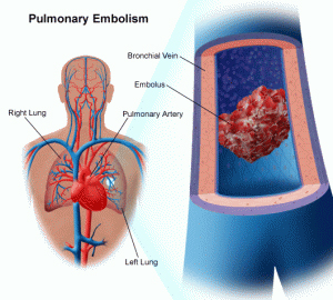
Pulmonary embolism is the obstruction of one or more pulmonary artery by a thrombus originating from somewhere. (Brunner and Suddarth)
Pulmonary embolism (PE) is a blockage of the main artery of the lung or one of its branches by a substance That has Travelled from elsewhere in the body through the bloodstream (embolism). (Wikipedia)

Causes
Most cases of pulmonary embolism according to Brunner and Suddarth caused by
1. Blood clot
2. Air bubble
3. Fat
4. Tumor cells

pulmonary artery, pulmonary fibrosis, pulmonary edema, signs pulmonary embolism, symptoms pulmonary embolism, pulmonary embolism definition, pulmonary embolism causes, pulmonary embolism cancer, pulmonary embolism ecg, pulmonary hypertension, treatment pulmonary embolism, acute pulmonary embolism, pulmonary embolism diagnosis, blood clot

Clinical Manifestations
The symptoms of pulmonary embolism depends on the size of the thrombus and the area of ​​the pulmonary artery is clogged by a thrombus. The symptoms may be nonspecific. Chest pain is the most common symptom and usually has a sudden onset and is pleuritic. Subternal sometimes can and can mimic angina pectoris or myocardial infarction. Dyspnea is the second most common symptom is followed by tachypnea, tachycardia, nervousness, cough, diaphoresis, hemoptysis, and syncope.
Massive embolism of the pulmonary artery bifurcation clog could cause actual dyspnea, substernal pain sudden, rapid and weak pulse, shock, syncope and sudden death.
Multiple small embolism can get stuck in the terminal pulmonary arteries, resulting in multiple small infarcts in the lungs. Clinical picture may resemble bronchopneumonia or heart failure. (Brunner and Suddarth)

Pathophysiology
When the thrombus blocking some or all pulmonary arteries, enlarged alveolar space loss due to the area, although they keep getting ventilation, receive little or no blood flow at all. In addition a number of substances that are released from the clot and cause the blood vessels constrict bronchiolus. This reaction in seimbangi ventilation perfusion imbalance, resulting in decreased O2 and increased CO2 levels.
Hemodynamic consequence is an increase in pulmonary vascular resistance due to decrease in size of pulmonary vascular net-nets., Causing an increase in pulmonary artery pressure and ultimately improve the working right ventricle to maintain pulmonary blood flow. When the right ventricle needs exceed the capacity, it will happen right ventricular failure that leads to a decrease in systemic blood pressure and shock. (Brunner and Suddarth)

Diagnostic Examination
Diagnostic examination of pulmonary embolism according to Brunner and Suddarth, are:
1. Chest X-rays
Chest radiograph in pulmonary embolism is usually normal but may shows a pneumo-constriction, infiltrates, atelectasis, elevation diagfragma the position of the sick, or dilation of the arteries and ​​pleural effusion.
2. ECG
ECG usually shows sinus tachycardia, atrial flutter or fibrillation and right axis deviation possible, or right ventricular strain.
3. Impedance Plethysmography
Plethysmography impedance performed to determine the presence of deep venous thrombosis.
4. Arterial blood gas
Arterial blood gases on pulmonary embolism may show hypoxemia and hipokapnea.

Investigations
Diagnostic examination that is often found on client pulmonary embolism as proposed by Doenges, M. E. (2000) the following:

  1. Lung scan (ventilation / perfusion scan) can show abnormal perfusion pattern in the area of ​​ventilation or absence of ventilation and perfusion.
  2. Pulmonary angiography: the presence of defects or cutoff in the absence of arterial blood in the distal blood flow.
  3. Chest X-ray: often normal (especially in the subacute state), but may show a shadow of blood clots, damage to blood vessels, elevation of the diaphragm on the affected area of ​​pleural effusion, infiltration consolidation.
  4.  ABGs: PaO2 may indicate a decrease, Paco, (hypoxemia, hipokapnea) and elevation of pH (respiratory alkalosis), especially if severe pulmonary obstruction.
  5. Complete blood: may show increased HT (hemoconcentration) increased red blood cell polistemia.
  6.  ECG : May be normal or show changes in the right ventricle that identify disorders, such as changes in ST segment or T waves, axis deviation / right bundle branch block (RBBB), tachycardia and dysrhythmias often occur.


Complication
Complications resulting from pulmonary embolism are:
1. Respiratory failure,
2. Acute right heart failure, and
3. Hypertension

Medical Management
According to Brunner and Suddarth (2001) The goal of treatment is to destroy (lyse) embolism pmbentukan existing and prevent new ones. Treatment of pulmonary embolism can include a variety of modalities:

  1. Anticoagulant therapy. Anticoagulation therapy include heparin, warfarin sodium has traditionally become the primary method to cope with acute deep vein thrombosis and pulmonary embolism.
  2. Thrombolytic therapy. Thrombolytic therapy include urokinase, streptokinase may also be used in dealing with pulmonary embolism, especially in highly disturbed paien. Thrombolytic therapy destroys thrombus or emboli faster and restore hemodynamic function of the pulmonary circulation is greater, because reducing pulmonary hypertension and improve perfusion, oxygenation, and cardiac output.
  3. Common action to improve the status of respiratory and vascular. Common actions undertaken to improve the status of respiratory and vascular patients. Oxygen therapy is given to correct hypoxia and to eliminate pulmonary vascular vasoconstriction and pulmonary hypertension and reduced.
  4. Surgical intervention.  Surgical intervention is carried out embolectomy lung, but embolectomy may be indicated in the following conditions:

a. if patients had persistent hypotension, shock, and severe heat
b. if very high pulmonary artery pressure
c. if the angiogram showed obstruction of the pulmonary blood vessels.
Embolectomy pulmonary require thoracotomy with cardiac pulmonary bypass technique

Tags : pulmonary artery, pulmonary fibrosis, pulmonary edema, signs pulmonary embolism, symptoms pulmonary embolism, pulmonary embolism definition, pulmonary embolism causes, pulmonary embolism cancer, pulmonary embolism ecg, pulmonary hypertension, treatment pulmonary embolism, acute pulmonary embolism, pulmonary embolism diagnosis, blood clot

Radiological Abnormalities of Atelectasis

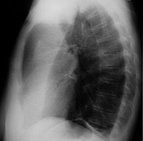
When the whole lung is deflated, there will be a homogeneous shadow on the sides of it, to heart and went to the department's trachea and the diaphragm lifted. When only one lobe of the atelaktasis caused by bronchial obstruction, may appear two characteristic abnormalities. The first abnormality is a shadow of a homogeneous than the deflated lobe itself, which will occupy a smaller space than when it develops at all.
Atelectasis treatment, atelectasis therapy, atelectasis Prevention, atelectasis antibiotics, Atelectasis Postural drainage
A right upper lobe collapse would appear as an opaque area at the top, with a limit that is strictly concave beneath the clavicle near the fissures caused by the raised horizontalis.

The left upper lobe usually includes Lingula when deflated, and the resulting image is less firm without the firm lower limit. But in the lateral projection would seem a tongue-shaped shadow with a peak near the diaphragm; anteriorly, it may be up to the sternum, or may be separated by a translucent area of ​​the lungs caused by a slip right next to them and the sternum posterior shadow It has a clear boundary with concave boundary caused by large fissures are pushed to the front.

A middle lobe will cause a shadow that is not firmly on the anterior projection, but may blur the line than the right heart, in the lateral projection it will appear as a ribbon-shaped shadow extending from the hilum to the angle of the sterno-diafragmatikus. Strict upper limits established by the nearest horizontalis fissures, whereas the concave rear boundary by a major fissure is pushed forward.

Lower lobe is deflated causing a triangular-shaped shadow, with the lateral border of the firm that ran downward and outward from the hilum to the diaphragm. Therefore it is usually located behind the heart shadow, he can only be seen when the radiograph is good. On the lateral projection image may be blurred at all, but its presence usually gives three images; thoracic vertebrae at the bottom will look more gray than black than the vertebrae next to the middle; the posterior than the shadow of the left diaphragm will not be seen; and finally, vertebrae in the back area below the heart shadow will be less black than the translucent area behind the sternum.

The symptoms of the other characteristics are the consequence rather than vascular shadows have become blurred in the general opacity than lobes that do not contain air, while the shadows of blood vessels in the other lobe is more dispersed because it fills a larger volume. Hilar blood vessels on the affected side will show a disease and not a lateral convexity concafitas as in the normal state at the place where the group rather than the upper lobe artery basal met in addition, the hilum will be smaller than on the other side, while blood vessels of the lungs will be more dispersed, so per unit area will look much less than on the other side (normal). Only there will be little or no relative translucency, because of capillary flow increases, Only there will be little or no relative translucency, because of capillary flow increases, whereas the tracheal pressure or elevation of the diaphragm and heart are usually a little switch only slightly in the direction of the deflated lobe of the collapse than the lower lobe, or more often as not at all on the upper lobe collapse rather than

Prevention & Treatment of Atelectasis

Prevention
Atelectasis treatment, atelectasis therapy, atelectasis Prevention, atelectasis antibiotics, Atelectasis Postural drainage
Treatment of atelectasis based on the etiology of the disease. However, prevention is the most important factor. The fundamental framework of therapy is early mobilization and frequent position changes on the client or the client postoperative bedrest. Breath in an orderly important because the client is generally a decline in the influence of anesthesia awareness, decreased mobility, and pain. Mucolytic bronchodilator and, if indicated, and chest physiotherapy will greatly help, adequate ventilation can be increased primarily to changes in position, effective coughing, deep breathing or incentive spirometry.
There are several ways you can do to prevent the occurrence of atelectasis:
  1. After undergoing surgery, the patient should be encouraged to breathe deeply, cough regularly and return to activity as quickly as possible.
  2. Although smokers have a greater risk, but this risk can be lowered by quitting smoking in 6-8 weeks before surgery.
  3. A person with chest abnormality or neurological conditions that cause shallow breathing in the long term, it might be better to use mechanical aids to assist breathing. This machine will produce continuous pressure to the lungs so that even at the end of a respiratory, respiratory tract can not be shrunk
  4. Encourage the client to breath deeply and effectively to prevent the blunting of secretion and to issue eksidat.
  5. Change patient position with frequent and regular basis, especially from the supine position to an upright position, to improve ventilation and prevent the accumulation of secretions.
  6. Increase chest expansion during breathing meeting for the deployment of air in the lungs as a whole.
  7. Give medications or sedatives wisely to prevent respiratory depression.
  8. Apply suction to remove secretions tracheobron chiolar.
  9. Perform postural drainage and chest percussion.
  10. Encourage activity or early ambulation.
  11. Teach proper technique insensif sporometri.

Treatment
The goal of treatment is to remove phlegm from the lungs and re-develop the affected lung tissue.
Actions are wont to do:
  • Lying on the side of healthy lungs so that the lungs are exposed again to inflate
  • Eliminate the stoppage, either by bronchoscopy or other procedures
  • Practice deep breathing (incentive spirometry)
  • Percussion (patting her) chest to dilute sputum
  • Postural drainage
  • Antibiotics are given for all infections
  • Treatment of tumors or other circumstances.
  • In certain cases, if the infection is persistent or recurrent, difficult or cause bleeding, it is usually part of the affected lung may need to be removed

Once the blockage is removed, usually gradually deflated lung will re-inflate, with or without the formation of scar tissue or other damage.
Bronchoscopy examination should be done immediately, if atelectasis occurs due to blockage of foreign objects. Provision of oxygenation should be given to patients with breathlessness and cyanosis. Symptomatic therapy is usually given as anti-tightness, bronchodilators, antibiotics and corticosteroids. Fisioterafi is unbelievably useful as position changes, massage, breathing exercises are very helpful in redeveloping the deflated lung.
In chronic infection that is usually done a more thorough bacteriological examination and lobectomy should not be done unless chronic infection and involves part of a healthy lung or has occurred bronchiectasis on a fairly wide area.

Tags : Atelectasis treatment, atelectasis therapy, Atelectasis Prevention, atelectasis antibiotics, Atelectasis Postural drainage

Related Posts Plugin for WordPress, Blogger...
Ping your blog, website, or RSS feed for Free TopOfBlogs

Followers