Definition | Treatment | Management of Disease | Diagnosis | Symptoms | Etiology | Pathophysiology | Procedure

 
Showing posts with label Treatment. Show all posts

Benefits and Risk of Asthma Drugs

asthma treatment, asthma medications, asthma therapy, Nebulizer therapy, Beta-agonist drug therapy, Risk of Asthma Drugs, Benefits of Asthma Drugs
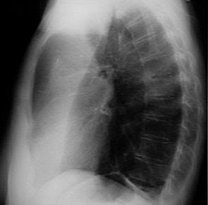
Talk about the asthma medication, not separated from the wide selection of types of drugs available. Starting from the class of drugs, intended use, as well as dosage forms. Different classes of drugs will show different effects. Different effects will affect the intended use, whether the drugs used to prevent or to cope with current asthma relapse. While the dosage form influencing onset (time taken from drug to drug consumption have an effect) and the effectiveness of medications so usually adjust to the goals of treatment and the patient's condition. But keep in mind the drug in addition to having the benefit, of course not separated from the risk of side effects.
Class of steroids. Examples of drugs known as steroids include budesonide, dexamethasone and beclometason. First-line drugs in asthma therapy is commonly used for the purpose of preventing a recurrence of asthma. But can also to address the current state of asthma relapse. In preventive therapy that requires patients take medication regularly should use inhalation dosage form, or better known as the metered dose inhaler (MDI). The use of inhaled has a faster onset compared with the use of oral (taken drugs thus bypassing the gastrointestinal tract). Side effects can be minimized because the drug only works around the respiratory tract. Regarding the issue of children's growth disorders and the onset of osteoporosis due to steroid use over and over, there are no facts for asthma drugs used in inhalation dosage forms. In addition to the inhalation dosage form, still it was likely to receive the steroid drug in oral dosage forms. Side effects of drugs known as steroids, among others, increasing pressure and blood sugar levels, so the use of steroids in people with hypertension and diabetes mellitus (DM) needs special attention. These drugs also have effects as steroids imunosupressan that can lower immunity. So you should still maintain the condition and stamina during its use. While the use of steroids to pregnant and lactating women is safe as long as the drug is given on the recommendation of doctors. Even before giving birth is often performed intravenous injections of drugs known as steroids to prevent asthma relapse during childbirth. Noteworthy is the time patients receive preventative therapy that requires the use of steroids on a regular basis. During steroid therapy received from outside bodies / exogenous system resulting in endogenous (hormones) in the body does not produce steroids. Therefore, the use of steroids should not be stopped abruptly, and the dose should be lowered slowly to give time to the endogenous system to get back to work producing steroids.
To overcome the acute attack, the drug class of beta-agonists such as salbutamol to be first-line drugs that work as bronchodilators (relaxing the bronchi). These drugs were already widely available in the form of inhalation so that the work is more effective in treating acute attacks. In an emergency where the patient has severe breathing difficulties that are used in nebulizer drug delivery methods. Nebulizer is a method of curing such a drug given to patients so that drugs can enter the respiratory tract even in difficult breathing. Unfortunately not all health facilities have the tools nebulizer because it is relatively expensive. In addition to the use of short-acting, there are also beta-agonist class of drugs that works long-acting, such as salmeterol or formeterol, onset and duration of which has a longer effect than salbutamol. Usually for the prevention of recurrence of asthma therapy. Side effects of beta-agonist group is quite diverse as: tremors / trembling of the hands, headache, hypokalemia (potassium deficiency), and tachycardia (accelerated heartbeat). However, these side effects do not always happen every time the use of drugs. Side effects appear or not depending on the clinical condition of each individual. If the beta-agonist drugs used in excessive long-term and may decrease its effectiveness. This is because the occurrence of drug receptor desensitization, so that the receptors become less sensitive. Therefore need larger doses to obtain the same effect. For that doctors will consider the most appropriate dose for patients according to clinical circumstances.
Beta-agonist drug therapy is sometimes combined with anticholinergic drug classes to achieve a better effect. Same with beta agonists, anticholinergic drugs such as ipratropium bromide group working with bronchial relaxing. Commonly used to treat acute attacks. Side effects that arise include: dry mouth, drowsiness, and impaired vision. Primarily on the use of inhalation technique in which patients perform less precise spraying. In a moment the eye can become blurred. Therefore, patients should know the proper technique of using inhaled eg by asking your doctor or pharmacist. One more familiar drug in the treatment of asthma, namely theophylline. Theophylline drugs classified as 'old' in the sense already used for therapy for a long time. Theophylline has a range of therapeutic dose and toxic dose is narrow. It can be dangerous if patients take excessive doses. Theophylline poisoning symptoms include: insomnia, headache, nausea, and tachycardia. Therefore at this time of theophylline has a lot left in asthma therapy. But sometimes still used for example in an emergency, theophylline administered by injecting in the form of aminophylline. Use of theophylline is considered because the price is economical. Theophylline was still there as one of the active ingredients-the-counter asthma medication. In conclusion asthma medications are quite safe. Recommended for use inhalation, because the effect is more rapid, appropriate target because it directly into the respiratory tract, side effects were minimal when compared to oral use so it is quite safe. And the proper technique of using inhaled greatly affect the success of therapy

Tags: asthma treatment, asthma medications, asthma therapy, Nebulizer therapy, Beta-agonist drug therapy, Risk of Asthma Drugs, Benefits of Asthma Drugs

Prevention & Treatment of Atelectasis

Prevention
Atelectasis treatment, atelectasis therapy, atelectasis Prevention, atelectasis antibiotics, Atelectasis Postural drainage
Treatment of atelectasis based on the etiology of the disease. However, prevention is the most important factor. The fundamental framework of therapy is early mobilization and frequent position changes on the client or the client postoperative bedrest. Breath in an orderly important because the client is generally a decline in the influence of anesthesia awareness, decreased mobility, and pain. Mucolytic bronchodilator and, if indicated, and chest physiotherapy will greatly help, adequate ventilation can be increased primarily to changes in position, effective coughing, deep breathing or incentive spirometry.
There are several ways you can do to prevent the occurrence of atelectasis:
  1. After undergoing surgery, the patient should be encouraged to breathe deeply, cough regularly and return to activity as quickly as possible.
  2. Although smokers have a greater risk, but this risk can be lowered by quitting smoking in 6-8 weeks before surgery.
  3. A person with chest abnormality or neurological conditions that cause shallow breathing in the long term, it might be better to use mechanical aids to assist breathing. This machine will produce continuous pressure to the lungs so that even at the end of a respiratory, respiratory tract can not be shrunk
  4. Encourage the client to breath deeply and effectively to prevent the blunting of secretion and to issue eksidat.
  5. Change patient position with frequent and regular basis, especially from the supine position to an upright position, to improve ventilation and prevent the accumulation of secretions.
  6. Increase chest expansion during breathing meeting for the deployment of air in the lungs as a whole.
  7. Give medications or sedatives wisely to prevent respiratory depression.
  8. Apply suction to remove secretions tracheobron chiolar.
  9. Perform postural drainage and chest percussion.
  10. Encourage activity or early ambulation.
  11. Teach proper technique insensif sporometri.

Treatment
The goal of treatment is to remove phlegm from the lungs and re-develop the affected lung tissue.
Actions are wont to do:
  • Lying on the side of healthy lungs so that the lungs are exposed again to inflate
  • Eliminate the stoppage, either by bronchoscopy or other procedures
  • Practice deep breathing (incentive spirometry)
  • Percussion (patting her) chest to dilute sputum
  • Postural drainage
  • Antibiotics are given for all infections
  • Treatment of tumors or other circumstances.
  • In certain cases, if the infection is persistent or recurrent, difficult or cause bleeding, it is usually part of the affected lung may need to be removed

Once the blockage is removed, usually gradually deflated lung will re-inflate, with or without the formation of scar tissue or other damage.
Bronchoscopy examination should be done immediately, if atelectasis occurs due to blockage of foreign objects. Provision of oxygenation should be given to patients with breathlessness and cyanosis. Symptomatic therapy is usually given as anti-tightness, bronchodilators, antibiotics and corticosteroids. Fisioterafi is unbelievably useful as position changes, massage, breathing exercises are very helpful in redeveloping the deflated lung.
In chronic infection that is usually done a more thorough bacteriological examination and lobectomy should not be done unless chronic infection and involves part of a healthy lung or has occurred bronchiectasis on a fairly wide area.

Tags : Atelectasis treatment, atelectasis therapy, Atelectasis Prevention, atelectasis antibiotics, Atelectasis Postural drainage

Management of COPD

The goal of COPD management are:
  1. Touch ups the ability of people with symptoms mengatasiu not only in the acute phase, but also the chronic phase.
  2. Improving the ability of the patient in carrying out daily activities.
  3. Reducing the rate of progression of disease if the disease can be detected early.


Management of COPD in the elderly are as follows:
  1. Etiological factors negate / precipitation, for example immediately stop smoking, avoid air pollution.
  2. Cleaning the bronchial secretions to help in various ways.
  3. Eradicate the infection with antimicrobial. In the absence of antimicrobial infections need not be given. Provision of appropriate antimicrobial should be in accordance with the germs that cause infections according to the results of sensitivity testing or empirical treatment.
  4. Overcome bronchospasm with bronchodilator drugs. The use of corticosteroids to resolve the inflammatory process (bronchospasm) is still controversial.
  5. Symptomatic treatment.
  6. Treatment of the complications that arise.
  7. Oxygen treatment, for those who need. Oxygen should be administered with a slow flow of 1-2 liters / minute.
  8. Rehabilitation actions which include:
  • Physiotherapy, mainly aims to help the expenditure of bronchial secretions.
  • Breathing exercises, to train the patient in order to make the most effective breathing.
  • Exercise with weights oalh particular sport, with the aim to restore physical fitness.
  • Vocational guidance, the work done against the patient can re-do their previous occupation.
  • Psychosocial management, primarily intended for patient self-adjustment to her illness.

 COPD (Chronic Obstructive Pulmonary Disease)

Tags : Management of COPD, COPD treatment, The goal of COPD management,

Asbestosis

Asbestosis is a respiratory disease caused by inhaling asbestos fibers, where the lungs extensive scar tissue is formed.

Asbestos minerals consist of silicate fibers with different chemical composition. If inhaled, asbestos fibers settle in the lungs, causing scarring.
Inhaling asbestos can also cause thickening of the pleura (the membrane lining the lungs).

Causes
Inhaling asbestos fibers can cause scarring (fibrosis) in the lungs.
Lung tissue fibrosis that forms can not inflate and deflate properly. Severity of the disease depends on the duration of exposure and the number of fibers inhaled.

mesothelioma asbestosis, mesothelioma, asbestosis cancer,  asbestosis disease, symptoms of asbestosis, asbestosis lung cancer, asbestosis exposure, what is asbestosis, asbestos exposure, asbestos symptoms, asbestosis causes, asbestosis compensation, asbestosis exposure, asbestosis pulmonar, asbestosis treatment, pulmonary asbestosis

Exposure to asbestos can be found in the mining and milling, construction and other industries.
Exposure to asbestos workers' families can also occur from particles brought home on the clothing of workers.

Diseases caused by asbestos include:
Mesothelioma can occur within 20-40 years after exposure.

Cigarette smoking causes increased risk of diseases caused by asbestos.
The number of events is equal to 4 among 10,000 people.

Symptoms
Asbestosis Symptoms appear gradually and emerging only after the formation of scar tissue in large quantities and the lungs lose their elasticity.

The first symptoms are shortness of breath and decreased ability to mild exercise.
About 15% of patients, will experience severe shortness of breath and respiratory failure.

Heavy smokers with chronic bronchitis and asbestosis, will suffer from coughing and wheezing.
Inhaling asbestos fibers can sometimes lead to accumulation of fluid in the space between the membranes lining the lungs. Although rare, asbestos can also cause tumors in the pleura, called mesothelioma, or the lining of the abdomen called peritoneal mesothelioma.

Mesothelioma caused by asbestos is malignant and can not be cured.
Mesothelioma generally arise stelah krokidolit exposed, one of four types of asbestos.
Amosit, other types, also cause mesothelioma.
Krisotil may not cause mesothelioma, but is sometimes tainted by tremolit that can cause mesothelioma.
Mesothelioma usually occur after exposure for 30-40 years.

Lung cancer will occur in patients with asbestosis who also smoke, especially those who smoked more than 1 (one) pack a day.

Other symptoms that may be found:
- Cough
- Tightness in chest
- Chest pain
- Nail abnormalities or clubbing fingers (fingers form that resembles a drum percussion).

Diagnosis
On physical examination using a stethoscope, you hear a voice crackles
To confirm the diagnosis, usually performed the following checks:
  • Chest radiograph
  • Lung function tests
  • CT scan of the lung.

Treatment
Supportive treatment to overcome the symptoms is to remove mucus / phlegm from the lungs through the procedure postural drainage, chest percussion and vibration.
Given drugs to thin the mucus spray.
May need to be given oxygen, either through a facemask (mask) or through a plastic tube that is placed in the nostrils.

Sometimes performed lung transplantation.
Fatal mesothelioma, chemotherapy is not very useful and removal of the tumor does not cure cancer.

Prevention
Asbestosis can be prevented by reducing levels of asbestos fibers and dust in the working environment.
Because the industry that uses asbestos dust control has been done, now suffering from asbestosis fewer, but still mesothelioma occur in people who have been exposed 40 years ago.

To reduce the risk of lung cancer, to workers dealing with asbestos, it is recommended to quit smoking.

Tags : Asbestosis definition, symptoms asbestosis, mesothelioma asbestosis, mesothelioma, asbestosis cancer,  asbestosis disease, symptoms of asbestosis, asbestosis lung cancer, asbestosis exposure, what is asbestosis, asbestos exposure, asbestos symptoms, asbestosis causes, asbestosis compensation, asbestosis exposure, asbestosis pulmonar, asbestosis treatment, pulmonary asbestosis

Management of Occupational asthma

To prevent the occurrence of occupational asthma is a medical examination prior to employment, use of protective equipment, monitoring air pollutants in the working environment is highly recommended. If occupational asthma has occurred, then transfer to the outside of the work environment is important. If for some reason can not be moved then it must be the prevention and monitoring of lung function decline.

Periodic evaluation of lung function in workers who already suffer from occupational asthma are needed to prevent disability. Clinical asthma will persist until a few years even though the worker has been out of work environment.


Medical treatment in patients with occupational asthma such as bronchial asthma in general:

  1. Theophylline, a bronchodilator and may suppress neutrophil chemotactic factor. Effectiveness of the two functions above depend on the serum levels of theophylline.
  2. Beta agonists, bronchodilators are the most good for the treatment of occupational asthma in comparison with cholinergic antagonists (ipratropium bromid).
  3. The combination of beta-agonists with ipratropium bromid improve lung function is better than just a beta agonist alone.
  4. Corticosteroids, from various studies is known to prevent the bronchoconstriction caused by bronchial provocation with allergens. It will also improve lung function, decrease exacerbations and airway hiperesponsivitas and will ultimately improve the quality of life.

Lung Cancer Treatment

Bronchial benign tumors are usually removed surgically because they can clog the bronchi and long may become malignant.
Sometimes performed surgery on the cancer other than small cell carcinoma that has not spread. Approximately 10-35% of cancers can be removed surgically, but surgery does not always bring healing.

Approximately 25-40% of patients and isolated tumor grows slowly, has a life expectancy of up to 5 years after his illness was diagnosed. Patients should perform routine checks for lung cancer recurrence in 6-12% of patients who had undergone surgery.

Before surgery, performed lung function tests to determine whether the remaining lung can still perform its functions well or not. If the result is ugly, it is not possible to do surgery.
Surgery is not necessary if:
- The cancer has spread beyond the lung
- Cancer is too close to the trachea
- Patients have serus circumstances (such as heart disease or lung disease is severe).


Radiation therapy performed in patients who can not undergo surgery because they have other serious illnesses.
The purpose of radiation is to slow cancer growth, not for healing. Radiation therapy also can reduce muscle pain, superior vena cava syndrome and suppression of the spinal cord. But radiation therapy can cause inflammation of the lungs (pneumonitis due to radiation), with symptoms such as cough, shortness of breath and fever. These symptoms can be reduced by corticosteroids (eg prednisone).

At the time of diagnosis, small cell carcinoma almost always has spread to other body parts, making it impossible to do surgery. These cancers are treated with chemotherapy, sometimes radiation therapy disetai.

Patients with lung cancer who experienced a lot of lung function decline. To reduce respiratory problems may be given oxygen therapy and drugs that dilate the airways (bronchodilators).

Lung Cancer Prevention

Tags : Lung Cancer, Lung Cancer Treatment, Treatment of lung cancer, treatment for cancer, treatment of cancer, chemotherapy treatment, cancer radiation treatment, radiation treatment, lung cancer chemotherapy, lung cancer survival

Management Of Pleural Effusion

Management Of pleural Effution :
  • Pleural fluid aspiration performed, to reduce the unpleasant taste or "discomfort" and shortness of breath.  It is advisable to gradually aspirations.Fluids released between 500 - 1000 cc. When taking too many and can quickly lead to pulmonary edema.
  • Incorporating intrapleura chemotherapy for malignancy (eg radioactive gold or Ytrium).
  • More often performed pleurodesis in the process of malignancy or in the frequent recurrence of effusion.
  • By using 500 mg tetracycline powder is dissolved in 50 cc of physiologic saline. Patients shake it so flat, then the liquid removed after clamped for 24 hours or given jodium powder or talc.
  • Pain that occurs because the administration of drugs above can be overcome with the analgesic. If need be given pethidine 100 mg i.m.
  • Provision of steroids combined with antituberculosis can absorb the pleural effusion caused by pulmonary tuberculosis quickly and reduce fibrosis.

 Management Of Pleural Effusion,Pleural effution, Pleural effussion, treatment pleural effusion,  Pleural effusion Procedure, left pleural effusion


Tags : Pleural effutionPleural effussion, treatment pleural effusion,  Pleural effusion Procedureleft pleural effusion

Special examination of Chronic bronchitis

Special examination :
  •  X-ray photo PA and left lateral chest
-  Typically show no abnormalities, except when it occurs cor pulmonale or emphysema.
-  It may also seem an increase in "bronchovascular markings".
  • Sputum: direct smear, Gram staining.


Please note: the existence of cells in sputum, whether the material is indeed contaminated with sputum or saliva from the oropharynx. Sputum can be identified because of alveoler macrophages (histiocytes). Macrophages alveoler this form round cells, large, with eccentric nuclei and oval or kidney-shaped near the edge of the cell.
Tues skuamus flat and very large and has a spherical nucleus in the middle. If there were> 25 epithelial cells in sputum skuamus, means contaminated with saliva or secretions from the oropharynx.
In chronic bronchitis who experience eksaserbrasi yellow sputum, bronchial ciliated epithelium that despite apparent in sputum neutrophil and looks.
At the time of remission decreased number of neutrophils, but macrophages alveoler increased, resulting in mucoid sputum and grayish-white color.
When sputum is contaminated, it should be repeated sputum collection or rinsed with water (washed sputum), a new Gram staining was made. Note the presence of neutrophils and bacteria. If the bacteria seemed gather very much and the same morphology, mean as a result of stasis, so sputum should be discarded. If it seems a variety of bacteria and many in the sputum, saliva possible, should be attempted again looking for new specimens. Look for the etiology of bronchial inflammation.
  • If the Gram-positive cocci in pairs or forming short chains means "Streptococcus pneumoniae" (diplococus).
  • If the Gram-positive cocci and form large clusters, meaning "Staphylococci".
  • Basil Gram-negative, such as haemophilus influenza, Seratia, Pseudomonas, Klebsiella, E. Colli, and others.

Guidelines for assessment of sputum on COPD:
  1. Gram staining of pus cells --> <1-5  inflammation (-) and germs (-); seed germ (+) --> not the cause of the disease.
  2. Gram staining (+) and seed (+) --> possible causes of infection.
  3.  Gram staining (-) and seed a little (+) --> sputum contaminated with saliva.
If the sputum culture results did not confirm the results of gram staining on sputum smears are true, then breeding considered untrustworthy. If the breeding is not the same as the results of gram staining, a new specimen should be made when antibiotics are used not deliver results.

Tests pulmonary physiology:
To determine the presence of airflow obstruction in chronic bronchitis, pulmonary physiology need to check:

  • FVC (Forced Vital Capacity)
  • FEV1 (Forced expiratory volume one second)
  • FEF 25-75% (Forced expiratory flow over the mid - 50% of the vital capacity) to detect obstruction in small airways.
  • PEFR (peak expiratory flow rate), only to find out the existence of a large obstruction in the airway, can be done in the clinic.
  • TLC (Total Lung Capacity), can only be examined at a hospital that has a complete facility in the form of "body plethysmograph or by helium dilution or nitrogen washout method technique".

In chronic bronchitis:
  • VC is normal or decreased (<80%)
  • FEV1 is normal or decreased (<80%)
  • FEV1 / FVC is always decreased (<75%)
  • FEF 25-75% was always decreased (<80%)
  • TLC normal / increased (N = 80-120%)
  • RV / TLC% is always increasing (N = 25-40%)

Pulmonary Physiology showed obstruction
  • ECG is necessary to know the existence of right heart hypertrophy, such as in cronic cor pulmonale.
  • Examination Arterial Blood Gases:
- PaO2 decreased to 70-80%, normal 80-100 mm Hg.
- PaCO2 normal / increased to 25-35%. Normal = PaCO2 = 35-45 mm Hg.
- pH is normal or decreased (when the state information). Normal pH = 7.35 to 7.45.
Tags : Bronchitischronic bronchitischronic coughchronic bronchitis cough, treatment chronic bronchitisManagement Chronic bronchitisbronchitis treatment,  acute bronchitis, Chronic Bronchitis Examination

Clinical Symtomps and Diagnosis Bronchiectasis

Clinical Symptoms of Bronchiectasis
  • Chronic productive cough, continuous or repetitive.
  • Coughing up blood, blood mixed sputum to massive coughing up blood.
  • acute exacerbation accompanied by heat.
  • Sputum mucoid, or purulent mukopurulen, when collected in a transparent glass look three layers: a layer of froth on top, the middle layer of mucus, pus and debris lining the bottom.
  • Shortness of breath, and breath sounds.
  • local wet crackles and settled.
  • Wheezing can be found.
  • Cachexia, cyanosis and clubbing in advanced cases.
diagnosis bronchiectasis, what is bronchiectasis, bronchiectasis COPD, bronchiectasis symptoms, bronchiectasis treatment, bronchiectasis definition, cystic bronchiectasis
Diagnosis
Diagnosis of bronchiectasis is established on the basic of:
  • Complaints and symptoms were found on physical examination.
  • Chest X-ray PA photo: normal in mild bronchiectasis. In severe cases, can seem "tram tracks" (two parallel lines like a tram tracks). The existence of the shadow ring when cut crosswise. If there is mucus plugging thick  --> linear density, or Y-shaped or V ("gloves-finger sign"). In cystic bronchiectasis, cystic cavities appear diameter of 3 cm sometimes appear water-fluid levels or nodules (when it was full).
  • Bronchography using contrast material, if planned for surgery. Picture looks cylindrical, varicose or cystic.
  • Examination of sputum (Gram and TTH), cultures of bacteria and fungi.
  • Antibiotic sensitivity tests.
  • Tests pulmonary physiology: VC and FEV1.
DIfferential Diagnosis

Complications of Bronchiectasis
Prognosis
Depending on the cause, location, and extent of abnormalities. With antibiotics and respiratory hygiene, improved prognosis.

Management of Bronchiectasis

Tags :diagnosis bronchiectasis, what is bronchiectasis, bronchiectasis COPD, bronchiectasis symptoms, bronchiectasis treatment, bronchiectasis definition, cystic bronchiectasis

Special Treatment of Bronchial Asthma

Special Treatment of Bronchial Asthma :
a. Mild asthma
Limitations:
Complaints arising tightness or cough less than two times a week, outside attacks asymptomatic sufferers. On physical activity may occur seranagn tightness or coughing which the period is short (<½ hours). Night asthma attacks rarely occur (<2 times a month). Pulmonary Physiology pasa asymptomatic state> / 80%, while the attacks may be decreased 20% or more.
asthma treatment, asthma bronchial treatment,  treatment of asthma, bronchial  asthma treatment
Treatment:
1. Beta 2 agonists: 2 sprays, may be repeated every 3-4 hours.
2. Cromolyn: can be added; before exposure to allergens, physical activity or other exposure.
b. Moderate Asthma
Limitations:
Complaints arise more frequently (> 1-2 times a week), that affect activity and sleep sufferers. Attacks can last several days. Sometimes required emergency treatment. Pulmonary physiology during asymptomatic approximately 60-80%, while time attack decreases to 20-30% or may be even tougher.
Treatment:
  • 2 times daily inhaled corticosteroids (400-800 ug / day) or cromolyn sprays 4 times a day 2. Coupled with the beta-2 agonist inhalation with a dose as needed up to 4 times a day. If more than 4 doses a day, need plus other drugs.
  • If still arise tightness, the dose of inhaled corticosteroids may be increased (up to 2000 mcg / day dose of> 1000 ug need strict supervision) and / or given oral theophylline (slow-release) and / or supplemented with beta-2 agonists by oral route.
  • Sometimes required oral corticosteroids (prednisone) for several days (40 mg / day in single or divided doses 2-4 doses) in a week, then lowered in a dose of next week.
c. Severe asthma
Limitations:
Complaints continued at any time with daily activities are limited. Frequent asthma attacks and often occur at night. Sometimes to require treatment in the emergency department or hospitalization.
Pulmonary Physiology day-to-day less than 60%, the attack can be decreased to 50%.
Treatment:
  • Inhaled corticosteroids 2-4 times a day 2-6 sprays (generally> 1000 ug / day) with or without cromolyn sprays 4 times a second and plus with beta-2 agonists. 
  • Beta-2 agonist inhalation, the dose is the same as in chronic asthma is. Can be given an additional extra 2-4 sprays in a day when required or given nebulizer.
  • To prevent asthma evening coupled with oral theophylline (slow-release) and / or beta-2 agonists by oral route.
  • Oral corticosteroids (prednisone) may be added to the dose and how as in chronic asthma is. Can be considered the provision in continuous with the minimal dose that gave the effect of a single repair 24/48 hour every morning.
d. Asthma and pregnancy
Uncontrolled asthma treatment can jeopardize health of mother and fetus. Will become more severe complications.
Treatment:
  • Must be optimal and should be given by inhalation.
  • Steroid injections can be administered, if necessary. (Risk to the fetus can be ignored).
Prognosis
Generally good, if diagnosis, treatment and prevention is made as early as possible with treatment adcquate.


Related Posts Plugin for WordPress, Blogger...
Ping your blog, website, or RSS feed for Free TopOfBlogs

Followers