Definition | Treatment | Management of Disease | Diagnosis | Symptoms | Etiology | Pathophysiology | Procedure


Management Of Hemothorax

Hemothorax patient death can be caused because of the large blood loss and the occurrence of respiratory failure.
Respiratory failure due to the large amount of blood in the pleural cavity pressure of lung tissue and reduced lung tissue that does ventilation.

Then treatment hemothorax as follows:
1. Emptying of blood from the pleural cavity.
Installed "chest tube" and is connected with the WSD system, this can accelerate the lung expands.
2. Stop the bleeding.

If the installation of WSD, the blood still does not stop, then considered for thoracotomy.
3. General state of repair.
Giving oxygen 2-4 liters / minute, the length adjusted to the clinical changes, better yet, if the monitored with blood gas analysis. Try to people with normal blood gases.
Giving blood transfusion: seen from a decrease in Hb.
As a benchmark can be used the following calculation, every 250 cc of blood (from patients with Hb 15 g%) can raise ¾ g% Hb.
Given with a normal drop of about 20-30 drops / minute and maintained not to an interruption in heart function or cause interference with the heart.
4. Others.
  • Antibiotics, carried out if there is secondary infection.
Antibiotics are used adjusted to the sensitivity test and culture. If the infecting organism is unclear, while the state of serious illnesses, then the patient can be given a "broad spectrum antibiotic", for example, ampicillin at a dose of 4 x 250 mg daily.
Also considered in case of pleural thickening decortication.

Tags : Management Hemothorax, Hemothorax procedure, Thoracotomy, hemothorax treatment, haemothorax, hemothorax
Hemothorax

Hemotorax is the presence of blood in the pleural cavity.

Pathophysiology and Etiology
  1. Sharp trauma or blunt trauma. This can occur when blunt trauma can cause fractures of the ribs, resulting in a torn intercostal blood vessels and also cause a tear in the lung tissue.
  2. A torn aortic aneurysm
  3. Complications due to drug administration on pulmonary infarction antikoagulansia
  4. In patients with abnormalities of "hemorrhagic diathese".
  5. Complications in thoracic surgery.
Clinical Symptoms


Symptoms and complaints hemothorax depending on the weight and severity of trauma. Patients may complain of shortness of breath, chest pain, until the shock and anemia.

Diagnosis
1. Anamnesa : A history of trauma to the chest, or after surgery.
2. Physical examination
  • Found such signs in the pleural effusion.
  • At the hospital hemitoraks reduced movement.
  • Hemithorax percussion on the sick and faint sounds on auscultation, breath sounds audible reduced or disappear altogether.
3. Chest X-ray photos : Radiological picture as in pleural effusion.
4. Laboratory.
After aspiration of the experiment, the liquid is carried out the examination as follows:
  • If the number of erythrocyte 5000-6000 / mm 3 --> "a Rosy tint".  If the number of red blood cells> 10,000 / mm 3 --> "serosanguinous". It is found in pleural effusion hemoragic.
  • If the fluid coming from complications hemoragis torasentesis, the liquid is centrifuged and the supernatant became clear. In contrast to the supernatant hemothorax still red.
  • It is said hemothorax tilapia Hb blood from the pleural cavity> 1 g / dl or if the hemoglobin derived from blood hemothorax half the price of capillary blood Hb.
Management Of Hemothorax

Complications
  1. Loss of blood.
  2. Respiratory failure.
Tags :definition hemothorax, hemothorax pleural effusion, hemothorax symptoms, tension hemothorax, hemothorax treatment, hemothorax chest tube, what is hemothorax, Management Hemothorax, Complications Hemothorax, hemothorax therapy, hemothorax procedure, Hemotorax, Haemothorax , Etiology Hemothorax, Pathophysiology Hemothorax

Management Of Pneumothorax

Outside the hospital.
  1. In light of spontaneous pneumothorax or pneumothorax simplex. Minimal or no complaints at all, are usually found by accident. The air in the pleural cavity will diresorbsi spontaneously. Because it does not require invasive measures.
  2. "Tension pneumothorax". Done in a sterile and carried out the stabbing in the sore area with a syringe the size of the largest. Stabbings in the space between the ribs into 2 in the front line of mid-clavicle. In young women (cosmetics) stabbings in the space between the ribs into 4 or 5 in the mid-axillary line. Then the needle tip covered with a sheet of thin rubber or thin plastic that can serve as a valve. Subsequently the patient was sent to hospital.
In the hospital.
  1. At the same place to do the installation of WSD, using trokar (troicar). It should be noted, that all actions undertaken SCARA sterile.
  2. WSD is removed, when the lung is expanding well and no complications after plastic hose clamped shut or 24 hours to prove that the pneumothorax was cured.
  3. If the patient is congested, it can be administered with high concentrations of oxygen and given to people with healthy lungs (before). In patients with COPD oxygen delivery must be careful.
  4. To treat pain may be given analgesics like-antalgin 3 x 1 tablet.
  5. In pneumothorax with severe COPD, is sometimes given strong analgesics such as pethidin 100 mg im or morphine 10 mg i.m. Physiotherapy should be given, because it could prevent sputum retention.
  6. If the lung development is rather slow, can be done with a suction pressure of 25-50 cm of water.
  7. In a recurrent pneumothorax (recurrent) do both pleural adhesions by using a material that can cause irritation or materials "scleroting agent".
  8. If there is a-Bronco-pleural fistula, it will be done eksterpasi operation.

Management of Empyema Thoracic

The principle of treatment in empyema:
1. Emptying of the pleural cavity of pus
2. Antibiotics
3. Closure of the pleural cavity
4. Causal treatment
5. Additional treatment.

1. Emptying of the pleural cavity.
a. Simple aspiration.
Performed repeatedly using a large needle hole. This method is good enough to remove most of the pus or fluid from acute empyema is still runny.
Losses such as these techniques often lead to "pocketed" empyema.
Ultrasound can be used to determine the localization of "pocketed" empyema.
b. Drainage is closed.
Installation "= closed thoracostomy tube drainage (WSD)".
Indications of this drain fitting, if the pus is very thick, pus is formed after 2 weeks and there has been piopneumotoraks.
Installation of the hose should not be too low, the diaphragm is usually raised because of empyema. Select a hose that is large enough.
If 3-4 weeks of no progress should be pursued by other means, such as in chronic empyema.
c. Installation of "open-drainage":
This action is done on a chronic empyema by cutting a piece of rib to create a "window". This method is chosen when dekortikasi not possible and should be done in a completely sterile condition.


2. Antibiotics.
Given the major cause of death due to sepsis, then antibiotics play an important role.
Antibiotics should be given immediately once the diagnosis is established and the diagnosis should adcquate. The selection of antibiotics based on results of Gram staining of pus smear.
Subsequent treatment depends on the culture results and sensitivity tests.
When germs that cause is unclear, can be used high doses of benzyl penicillin.
3. Closure of the pleural cavity.
When chronic empyema fails to show a response to the drainage hose, then do dekortikasi or torakoplasti.
If not handled properly will add to the long hospitalization.
4. Causal treatment.
Depending on the cause eg, amubiasis, tuberculosis, aktinomikosis.
Treated with specific drugs for each disease.
5. Additional treatment and physiotherapy.
Aiming to improve the general situation

Tags = Empyema treatment, Antibiotic for EmpyemaManagement Of empyema thoracic, management for empyema thoracic, empyema thoracic management, empyema thoracic treatment, treatment of empyema thoracic, empyema thoracic medical management, Management Of empyema thoracis, management for empyema thoracis, empyema thoracis management, empyema thoracis treatment, treatment of empyema thoracis, empyema thoracis medical management

Empyema Thoracic

Empyema Thoracic is the presence of pus in the cavity / pleural cavity.

Etiology of Empyema Thorasic
I. Derived from the lungs:
  1. Pneumonia
  2. Lung abscess
  3. The existence of bronchopleural fistula
  4. Bronchiectasis
  5. Pulmonary Tuberculosis
  6. Lung fungus.

II. From the extrapulmonary infection:
  1. Trauma of the brain
  2. Brain surgery
  3. Torasentesis
  4. Subfrenik abscess
  5. Due to amoebic liver abscess.

Bacteriology
  1. Staphylococcal piogenes, at all ages, often in children.
  2. Piogenes streptococcus.
  3. Gram-negative bacteria (Pseudomonas aeruginosa,, Klebsiela, Bakteroides, E. coli, Proteus mirabilis)
  4. Anaerobic bacteria.

Pathophysiology Of Empyema Thorasic
Due to pyogenic bacteria invasion into the pleura arising acute inflammation, followed by the formation of serous exudates. With the number of PMN cells either living or dead, as well as increased levels of protein, the fluid becomes cloudy and thick. Fouled fibrin will form pockets of pus to localize it.

Clinical Symptoms
Clinical Course
Divided into 2 stages, namely: acute and chronic.
1. Acute empyema:
Symptoms are similar to pneumonia, high fever, pleuritic pain when the stage is left in a few weeks there will be toxemia, anemia and clubbing. If pus is not issued soon will arise fistula bronkopleura and "empyema necessitatis".
2. Chronic empyema:
Strict boundary between acute and chronic hard set, called chronic empyema when running more than 3 months.
Patients complain of a weak body, health, the patient looked back, pale and no clubbing.

Diagnosis
Physical examination.
Found signs of fluid, accompanied by the movement of sick hemitoraks reduced. There was a faint sound on percussion. On auscultation, breath sounds decreased until it disappears in the hemitoraks sick.

Chest X-ray photo.
On chest PA X-ray and lateral images obtained an image of "opacity" that indicate the presence of fluid with or without pulmonary abnormalities.
In the event of fibrotoraks, trachea and mediastinum attracted to the side of the hospital and also looked pleural thickening.

Sure Diagnosis
Pleural aspiration would indicate the presence of pus in the pleural cavity.
Furthermore, the pus is used as material for examination: cytology, bacteriology fungi, amoeba, done culture (culture) and sensitivity to antibiotics.

Management of Empyema Thoracic


Complications of Empyema Thoracic
Complication that often arises is: bronchopleural fistula.
Other complications that may occur are: shock, sepsis, congestive heart failure and otitis media.


Tags : empyema thoracis, Definition Empyema, Empyema thoracic, Empyema symtomps, Empyema treatment, What is Empyema, Management Empyama, empyema treatment

Management of Bronchiectasis

A. Conservative:
1. Control of acute and chronic infection, mucus secretion, airway obstruction and complications, such as: coughing up blood, respiratory failure and cor pulmonale, in order to prolong life, improve quality of life and prevent disease progression.
2. Antibiotics if there is infection.
3. Chest physiotherapy and postural drainage with forced expiratory technique for removing secretions.
4. Aerosols with physiologic saline or beta agonists prior to chest physiotherapy may facilitate the release of sputum / secretions.
5. Bronchodilators to improve airflow, helping mukosilia clearance and physiotherapy improve outcomes.
6. Corticosteroids when there is severe bronchospasm (CPOD or Asthma Bronchiale).
bronchiectasis pneumonia, what is bronchiectasis, bronchiectasis copd, bronchiectasis symptoms, bronchiectasis treatment, bronchiectasis definition, cystic bronchiectasis, Management bronchiectasis, Procedure of bronchiectasis
B. Surgery:
Indications of surgery:

Tags : bronchiectasis pneumonia, what is bronchiectasis, bronchiectasis copd, bronchiectasis symptoms, bronchiectasis treatment, bronchiectasis definition, cystic bronchiectasis, Management bronchiectasis, Procedure of bronchiectasis

Clinical Symtomps and Diagnosis Bronchiectasis

Clinical Symptoms of Bronchiectasis
  • Chronic productive cough, continuous or repetitive.
  • Coughing up blood, blood mixed sputum to massive coughing up blood.
  • acute exacerbation accompanied by heat.
  • Sputum mucoid, or purulent mukopurulen, when collected in a transparent glass look three layers: a layer of froth on top, the middle layer of mucus, pus and debris lining the bottom.
  • Shortness of breath, and breath sounds.
  • local wet crackles and settled.
  • Wheezing can be found.
  • Cachexia, cyanosis and clubbing in advanced cases.
diagnosis bronchiectasis, what is bronchiectasis, bronchiectasis COPD, bronchiectasis symptoms, bronchiectasis treatment, bronchiectasis definition, cystic bronchiectasis
Diagnosis
Diagnosis of bronchiectasis is established on the basic of:
  • Complaints and symptoms were found on physical examination.
  • Chest X-ray PA photo: normal in mild bronchiectasis. In severe cases, can seem "tram tracks" (two parallel lines like a tram tracks). The existence of the shadow ring when cut crosswise. If there is mucus plugging thick  --> linear density, or Y-shaped or V ("gloves-finger sign"). In cystic bronchiectasis, cystic cavities appear diameter of 3 cm sometimes appear water-fluid levels or nodules (when it was full).
  • Bronchography using contrast material, if planned for surgery. Picture looks cylindrical, varicose or cystic.
  • Examination of sputum (Gram and TTH), cultures of bacteria and fungi.
  • Antibiotic sensitivity tests.
  • Tests pulmonary physiology: VC and FEV1.
DIfferential Diagnosis

Complications of Bronchiectasis
Prognosis
Depending on the cause, location, and extent of abnormalities. With antibiotics and respiratory hygiene, improved prognosis.

Management of Bronchiectasis

Tags :diagnosis bronchiectasis, what is bronchiectasis, bronchiectasis COPD, bronchiectasis symptoms, bronchiectasis treatment, bronchiectasis definition, cystic bronchiectasis

Bronchiectasis

Bronchiectasis is abnormal and permanent dilatation of bronchi and cartilaginous being, accompanied by destruction of muscle and elastic components of the walls.

Etiology and pathogenesis
A bronchial wall inflammation, causing damage and dilatation. Contributing factors are:

1. Infection: primary and secondary.
Primary or secondary infection, either repeatedly or continuously, by: bacteria, viruses, mycoplasma, and mycobacterial clearance mukosilia and will damage the airway epithelium.
In children is often caused by: measles, whooping cough, severe pneumonia or aspiration. Primary tuberculosis is often also leads to bronchiectasis.
bronchiectasis pneumonia, what is bronchiectasis, bronchiectasis copd, bronchiectasis symptoms, bronchiectasis treatment, bronchiectasis definition, cystic bronchiectasis
Endobronchial tuberculosis causing necrosis, focal stenosis due to endobronchial inflammation, enlarged lymph nodes give emphasis or endobronchial obstruction, and scarring of the parenchyma causes distortion of the airway.

2. Inhalation of toxic chemicals or materials imunoaktif.
Inhalation of toxic chemicals or materials imunoaktif or autoimmune reaction: anhydrous ammonia vapor, sulfur oxides, talc, cork, bakelite, and smoke. Aspiration of gastric contents resulting in inflammation and result in bronchiectasis, for example in the hiatal hernia, gastric motility disorders, divertikuli, trakeoesofageal fistula.

3. An abnormal immune response: a genetic or acquired.
Abnormal immune response: a genetic or "acquired".
Swollen lungs because of heroin or heroin poisoning can cause bronchiectasis, due to changes in immunological defense.
Bronchiectasis occurs in patients with ulserosa colitis, rheumatoid arthritis, Sjogren's syndrome, cutaneous vasculitis, Hashimoto's thyroiditis, pernicious anemia, "primary biliary cirrhosis," "celiac disease", and sarcoid, because of an abnormal immunological manifestations, which often form hiperresponsif or autoimmune response.
4. Mechanical factors.
Atelectasis or parenchymal fibrosis can lead to bronchiectasis. Atelectasis can cause bronchial dilatation is back to normal when the lungs expand again. Parenchymal fibrosis can lead to bronchial dilation through withdrawal.
Bronchial dilatation and distortion may interfere with clearance mukosilier, leads to accumulation of secretions, resulting in infection with bronchial wall damage elements --> bronchiectasis.

5. Inherited or congenital abnormality.
Deficiency of alpha-1-antitrypsin causes emphysema panasinar and diffuse bronchiectasis.

  • "Allergic bronchopulmonary aspergillosis" --> central or proximal bronchiectasis.
  • BEAP syndrome (bronchiectasis, eosinophilia, asthma, and pneumonitis).
  • Bronchiectasis is a picture that stands out in cystic fibrosis, in children and adults.
  • Kartegener syndrome (bronchiectasis, sinusitis, site inversus).
  • Young's syndrome: obstructive Azoospermia and lung abnormalities (bronchiectasis).
  • "Pulmonary sequestration".
  • Yellow nail syndrome.
  • William Syndrome - Campbell (deficiency of bronchial cartilage generation 4 through 8).
  • Swyer Yndrome - James and Mac Leod (hipertensi unilateral lung).
  • Marfan's syndrome.

Clinical Symtomps and Diagnosis Bronchiectasis

Management of Bronchiectasis

Tags : bronchiectasis pneumonia, what is bronchiectasis, bronchiectasis copd, bronchiectasis symptoms, bronchiectasis treatment, bronchiectasis definition, cystic bronchiectasis

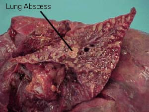
Lung Abscess

Lung abscess is a suppurative lesions accompanied by necrosis of tissue in it.

Etiology
Germs cause usually consist of a mixture of aerobic and anaerobic bacteria such as: peptokokus, peptostreptokokus, fusobakterium spp, bakteroides spp, which is the flora of oropharynx.
An abscess can occur because: aspiration, complications of pneumonia, lung trauma wounds are infected, and infected with pulmonary infarction or originating from empyema.
In the subsequent discussion only lung abscess caused by the aspiration to be described further.

Pathogenesis

Infection will easily arise when there predisposing factors, such as:
  1. A source of infection is an infection of the respiratory tract mouth, larynx tumors infected, infected bronchiectasis, and lung tumors are infected.
  2. The resistance of the airways that decrease, due to disorders such as laryngeal paralysis, a decreased patient consciousness, achalasia, esophageal carcinoma, and disorders expectorated.
  3. Mechanical airway obstruction due to aspiration of blood clots, pus, teeth, vomit, bronchial tumors.


Material that is inhaled into the lungs will lower lying (gravity-dependent segments). In the upright position will aspirate into the basal segment of inferior lobe, especially the right, but if aspiration occurs during sleep supine aspiratnya will go to the posterior segment of superior lobe and inferior lobe superior segment.
The process begins as a pneumonia, and if they do not receive adequate treatment, then the process will evolve into "necrotizing pneumonia" or a lung abscess.

Pathology
Cavity caused by tissue necrosis, surrounded by thick walls and surrounding lung inflammation. Usually have a relationship with bronchial cavities.

Clinical Symptoms
Cough, foul smelling sputum, fever, pleuritic pain, the agency added skinny, sweaty night. A typical course of the disease is chronic and slow (chronic and indolent) potential for the occurrence of sudden and serious complications such as brain abscess, coughing up blood profus, piopneumothorax.

Diagnosis
Diagnosis of lung abscess due to aspiration is made by:
  1. A history of aspiration, especially in sufferers with impaired consciousness, impaired swallowing. But the state of sleep often unconscious aspirations. Another predisposing condition for anaerobic infections.
  2. Typical clinical symptoms: a chronic course of the disease and indolent. Cough with purulent sputum stink.
  3. Abnormalities in one place in the lung in accordance with the position of the patient at the time of aspiration.

Physical examination
Found signs of consolidation and the sign of a cavity in the sore area.
Inspection: decreased respiratory movement
Palpation: fremitus conjecture elevated in diseased areas. Sometimes palpable pleural friction.
Percussion: sounds overcast condition at the regional hospital.
Auscultation: the diseased areas of bronchial breathing sounds. Accompanied by additional sound rough to smooth. Above the cavity will sound amforik.
Laboratory:
- Blood edge: leukocyte increase was 12,000 - 20.000/ml erythrocyte sedimentation rate increased anemia.
- Sputum: Gram staining obtained many PMNs, and bacteria of various types.
Chest X-ray photo: looking to the consolidation of part or one lobe in which there is "water fluid levels".

Differential Diagnosis of Lung abscess
  1. Pulmonary tuberculosis is usually not accompanied by "air fluid levels"
  2. Bronchogenic carcinoma who experienced necrosis. Cavity wall thick, not flat.
  3. Bulla or cysts that are infected are marked with thin walls, no surrounding inflammatory reaction.
  4. Pulmonary hematoma is characterized by a history of trauma, no signs of infection.
  5. Skwester experiencing lung abscess formation. No association with bronchial (bronchography).
  6. Pneumoconiosis are experiencing cavitation and marked by a "simple pneumoconiosis" around it.
  7. Hiatus hernia. In hiatus hernia, there are no symptoms from the lungs. Found the existence of retrosternal pain and burning sensation is felt in the pit of the stomach (heart burn). This pain will be worse if the patient bent.
Complications of Lung abscess
  1. Coughing blood profus
  2. Empyema or piopneumothorax
  3. Brain abscess
  4. Anemia,caHection and amyloidosis can occur in chronic disease.
Clikc Here Management Of  Lung Abscess

Prognosis
When treatment is appropriate, given the prognosis is not too late either.


Tags : abscess of lung, abscess in lunglung abscess treatment, lung abscess symptoms, pulmonary abscess, abscess on lung, Absces in lung, Absces in lung, Lung Abcess treatment, lung Abcess

Management of Lung abscess

1. General management:
Improving the general condition of patients with high-calorie high-protein diet and drink plenty of fluids.
a. Antibiotics.
  • Procaine penicillin G given 1.2 million units im every 12 hours + chloramphenicol 500 mg every 6 hours for 10-15 days. or
  • Procaine penicillin G 1.2 million units i.m. every 12 hours + Metronidazole 500 mg every 6 hours for 10-15 days. or
  • Clindamycin 600 mg every 8 hours for 10-15 days.

b. Postural drainage and physiotherapy.
The position of the body are arranged so that pus can come out by itself (due to gravity) or with the help of the physiotherapist.

2. Special Treatment:
a. Bronchoscopy
If pus is difficult exit, it is necessary to bronchoscopy to clear the airway and sucking pussy.
b. Surgery
When chemotherapy failed. A chronic abscess, cavity remains and sputum production remained there while the clinical symptoms are still present after adequate therapy for 6 weeks or the rest of extensive scar tissue that can interfere with lung physiology. This is all an indication of surgery.

Tags : Management Of Lung abscess, management for Lung abscess, Lung abscess management, community acquired Lung abscess, Lung abscess treatment, treatment of Lung abscess, Lung abscess medical management

Management of Bacterial Pneumonia

Indications of hospitalization is:
1. Patients with basic diseases
2. Patients with complications.

General management.
1. Correction of underlying abnormalities (underlying disease)
2. Bedrest
3. Symptomatic medications, administered only if necessary, such as:
  • 3 x 500 mg Paracetamol (in hyperpyrexia)
  • Morphine 10 mg s.c. (If there is pain severe).

4. Maintain fluid and electrolyte balance with the help of intravenous fluids, 5% dextrose, normal saline or Ringer's lactate.

5. Selection of anti-infective drugs.
Selection of antimicrobial drugs, should be based on sensitivity tests and sensitivity, but due to time and facilities greatly affect the success of this test, then giving more medicine based on empirical, as below:

Special Treatment:
1. Pneumococcal pneumonia,
Basic disease: elderly, Chronic Obstructive Pulmonary Disease (COPD), CHD, diabetes mellitus, alcoholism, post-influenza.
Clinical features: sudden illness, high fever, chills, pleuritic pain, cough productive with phlegm such as iron rust, herpes labialis.
Physical diagnostics: signs of consolidation.
Chest X photo: visible presence consolidated picture.
Laboratory:
- Blood banks: lekositosis 15,000 - 40.000/ml.
- Sputum: gram positive cocci which are lancet-shaped.
Treatment:
a. Antibiotics options:
Penicillin G from 0.6 to 1.2 million units i.m. every 12 hours, for 50-10 days.
b. Antibiotics alternatives:
Erythromycin 500 mg orally every 8 hours for 5-10 days or Tetracycline 500 mg orally every 8 hours for 5-10 days.

2. Staphylococcal pneumonia.
Basic disease: post-influenza, diabetes mellitus, malignancy, advanced age, use of drugs.
Clinical features: sudden onset of illness accompanied by high fever, chills, pleuritic pain and shortness of breath accompanied by cough productive with purulent sputum (with blood).
Physical diagnostics: look for signs of consolidation in one lobe or
several segments, and quickly arise necrosis, abscess and
pneumatokel.
Chest X photo: a picture of consolidation in one lobe or the
some segments.
Laboratory:
- Blood banks: leucocytes 15,000 - 35.000/ml
- Sputum: gram-positive cocci found that gather.
Treatment:
a. Antibiotic choice: cloxacillin 250-500 mg iv given every 6 hours for 5-10 days.
b. Alternative antibiotics: Cephalexin / Sefazolin 0.5 to 1 gram, 3 times a day, for 50-10 days.

3. Pneumonia klebsiela.
Basic diseaseChronic Obstructive Pulmonary Disease (COPD), alcoholics, elderly, diabetes mellitus
Clinical symptoms: sudden illness, high fever, chills, pleuritic pain, shortness of breath, productive cough with phlegm like jelly (currant jelly)
Physical diagnostics: according to the signs of consolidation, chest looks
more convex.
Chest X photo: picture looks consolidation with an enlarged volume
so that the fissure becomes convex (bulging).
Laboratory:
- Blood banks: leucocytes 15,000 - 40.000/ml.
- Sputum: gram-negative bacilli appear in the form of fat.
Treatment:
a. Antibiotic choice: Ampicillin 1000 mg i.v. given every 6 hours, plus Gentamicin 60-80 mg every 8 hours, for 50-10 days.
b. Alternative antibiotics: Cefotaxime 1 g i.v. every 6 hours for 5-10 days.

Tags : management of pneumonia, management for pneumonia, pneumonia management, community acquired pneumonia, pneumonia treatment, treatment of pneumonia, pneumonia medical management

Bacterial Pneumonia

Bacterial Pneumonia Is an acute infection of lung parenchyma caused by bacteria. Pneumonia is an infectious disease other than that often obtained in the community (community acquired pneumonia), he also often acquired in hospital (hospital acquired pneumonia = nosocomial pneumonia).
Pneumonia is the second difference, lies in the etiology and management.

Etiology
Bacterial pneumonia, can be basically caused by all kinds of bacteria, but most are caused by streptococcus pneumonia (80%), staphylococcus aureus, haemophilus influenza, pneumonia klebsiela germs while others are very rare.
Pneumonia, Bacterial Pneumonia,Pneumonia definition, pneumonia symptoms, symptoms of pneumonia, pneumonia treatment, bronchitis pneumonia, pneumonia contagious, walking pneumonia symptoms, what is pneumonia
Pathogenesis
The bacteria enter the lungs through:
  1. Aspiration of secretions from the oropharyngeal
  2. Inhalation of fine granules sputum (droplet)
  3. Channels of blood from an outside source of pulmonary infection (haematogenous).

Pathology
Germ that enters the alveoli causes an inflammatory reaction that can spread through the process of Kohn and airways to the surrounding lung parenchyma.
These inflammatory cells to satisfy one segment of one lobe extends into the forming process of consolidation. This process causes lung tissue become dense and resembles a heart so called hepatization.
Particularly for infections caused by Staphylococcus aureus, where the infection process begins in the bronchioles and accompanied by focal necrosis and abscess formation, then spread to peribronchial will facilitate the emergence of pneumatokel. Germ staphylococcus aureus, in its distribution can also reach the lung tissue through the septic emboli.


Clinical Symptoms
Characterized by the presence of acute infection and pulmonary consolidation.
Usually there are circumstances that underlie the onset of acute infection, such as: advanced age,Chronic Obstructive Pulmonary Disease (COPD), diabetes, alcoholism, post-influenza etc..

Acute infection is characterized by:
Sudden high fever with chills. Body temperature can reach 40 degrees Celsius or more and accompanied by general symptoms such as malaise, weakness, weight and appetite loss.

Pulmonary inflammation characterized by:
Chest pain (pleuritic) in place processes, shortness of breath, cough with purulent sputum are sometimes mixed with blood.

Diagnosis
Diagnosis of a bacterial pneumonia based on:
1. History of disease, signs of acute infection that usually there are basically disease (see clinical symptoms).
2. Signs of lung consolidation, and the sick lung area found abnormalities in the form:
Inspection: left pulmonary respiratory movements, because the patient sought
withstand the movement of the sore area with your hands or a pillow.
Palpation: fremitus touched up. Palpable presence of pleural friction.
Percussion: the sound of overcast condition in accordance with the lobes or the appropriate boundary
with an area of ​​the sore area.
Auscultation: breath sounds audible to the bronchial bronchovesikuler.
Sometimes the sound of extra breaths of crackles. Be accompanied
with positive bronchofoni and whispered voice.
3. Laboratory:
Peripheral blood: leucositosis 15,000 - 40.000/ml.
Sputum: macroscopic, purulent blood mixed with or without microscopically, the painting looks gram positive cocci gram and / or gram-negative bacilli along with many types of  Polimorfonukleus (PMN) leucocytes.
Chest X-ray photo: looks a gloom on one lobe and accompanied by a picture bronchogram air.

Differential Diagnosis
  • Non-bacterial pneumonia
  • Pulmonary tuberculosis, particularly tuberculosis pneumonia
  • Non-infectious pneumonia
  • Puffy Lung
  • Pulmonary infarction.

Complications

Prognosis
  • Community acquired pneumonia prognosis is good as long as not too late for treatment and receive adequate treatment.
  • Hospital acquired pneumonia prognosis is worse, because bacteria are often resistant to various drugs (multi resistant).

Tags: Pneumonia definition, pneumonia symptoms, symptoms of pneumonia, pneumonia treatment, bronchitis pneumonia, pneumonia contagious, walking pneumonia symptoms, what is pneumonia,

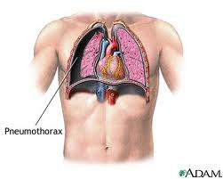
Pneumothorax (Collapsed lung)

Pneumothorax is the obtainment of air in the pleural cavity.
tension pneumothorax, spontaneous pneumothorax, spontaneous, pneumothorax symptoms, pneumothorax treatment , open pneumothorax, pneumothorax symptoms, atelectasis, symptoms of pneumothorax, pneumothorax definition, right pneumothorax, pneumothorax causes, pneumothorax, what is pneumothorax, Hemopneumothrax

Pathophysiology and Etiology
  1. Spontaneous pneumothorax due largely located superficial bullae rupture, and preceded by an increase in intra-pulmonary pressure, among others: cough hard or after blowing musical instruments, sneezing, straining, and others. The entry of air into the pleural cavity, through a tear in the visceral pleura. Bula congenital, predisposing especially in young males. Bula may also arise due to pulmonary tuberculosis, pneumoconiosis and bronchial obstruction.
  2. Traumatic pneumothorax can be caused by a "penetrating and non penetrating injury," either with or without rib fractures.
  3. "Surgical trauma" and "iatrogenic damage". Surgery can cause this type of pneumothorax.
  4. "Artificial pneumothorax" necessary for the treatment of hemoptysis in pulmonary tuberculosis as well as diagnostic measures for lung tumors.
  5. "Tension pneumothorax" due to "check valve" mecanism, so that air can enter but can not get out of the pleural cavity to the mediastinum due to a healthy one side driven.
  6. Open pneumothorax, occurs when the visceral pleura remains torn open so that the pressure in the pleural cavity with the outside air pressure.
  7. Closed pneumothorax, when ripping a hole closes after the incoming air is quite a lot. As a result the pressure in the pleural cavity is higher than the outside air pressure.
  8. Hemopneumothoraks, hidropneumothoraks and piopneumothoraks, when the pleural cavity contains blood, pleural fluid or pus clear.

Symtoms and Clinical Examination

  1. Sudden chest pain
  2. Sudden shortness of breath
  3. Respiratory failure and may be accompanied by cyanosis Puls.
Physical examination
  1. There is often a "Circulatory collapse" because of "Tension pneumothorax".
  2. In the percussion sound obtained hipersonor
  3. On auscultation, decreased breath sounds found on the side until the pain disappears.

Chest X-ray Pictures
1. On PA X-ray chest images visible edge of a collapsed lung in the form of a line. In pneumothorax parsialis the localization in the anterior or posterior, the perimeter of the lung may not be visible.
2. "Mediastinal shift" can be seen in the photograph Anatotomical Pathology or fluoroscopy when the patient inspiration or expiration, especially can occur in "Tension pneumothorax".

Differential Diagnosis
1. Pleurisy and pericarditis
2. Myocardial infarction and pulmonary embolism
3. Chronic bronchitis and emphysema
4. "Diaphragmatic herniae"
5. "Dissecting aneurysmae aortae".

Indication-specific treatment:
1. "Tension pneumothorax"
2. Pneumothorax accompanied by shortness of breath.
3. Bilateral pneumothorax
4. Large pneumothorax. When a line edge of a collapsed lung> 1 / 3 transverse diameter.
5. There appear to pleural fluid accumulation are aplenty
6. Pneumothorax as a complication of pulmonary diseases, such as supurativa pneumonia, tuberculosis.
7. Recurrent pneumothorax
8. Complications in the use of a ventilator.

Procedure and Management Of Pneumothorax

Complications
  1. "Tension pneumothorax" will berakhur fatal, if there is "Circulatory collapse".
  2. Respiratory failure.
  3. Hemopneumothrax
  4. Secondary infections
  5. Pleural thickening
  6. Atelectasis
  7. Recurring. 20% in simple or pneumothraks pneumothraks simplex and 50% in patients with COPD
  8. Mediastinal emphysema
  9. "Re-expansion pulmonary oedema".
Prognosis
  1. Good, if immediate relief and intensive treatment, especially regarding healthy young patients.
  2. Essentially dependent disease or "underlying disease", is very dangerous when the patient with COPD.

Reference

  1. Crofton J, Douglas A. Respiratory Disease, 3rd edition . Blackwell Scientific Publications, Singapore, 1984, pp. 541 – 546.
  2. Graham K, Crompton. Diagnosis and Management of Respiratory Diseases. Blackwell Scientific Publications. Oxford-London- Edinburg 1980, p. 150 – 163.
  3. Sol Katz. Spontaneus Pneumothorax In : Respiratory Emergency. Ed. By Moser KM, Spragg, R.G, 2nd edition. The CV Mosby Company, London, 1982, p. 176 – 193.

Tags : tension pneumothorax, spontaneous pneumothorax, spontaneous, pneumothorax symptoms, pneumothorax treatment , open pneumothorax, pneumothorax symptoms, symptoms of pneumothorax, pneumothorax definition, right pneumothorax, pneumothorax causes, pneumothorax, what is pneumothorax, Hemopneumothrax

Special Treatment of Bronchial Asthma

Special Treatment of Bronchial Asthma :
a. Mild asthma
Limitations:
Complaints arising tightness or cough less than two times a week, outside attacks asymptomatic sufferers. On physical activity may occur seranagn tightness or coughing which the period is short (<½ hours). Night asthma attacks rarely occur (<2 times a month). Pulmonary Physiology pasa asymptomatic state> / 80%, while the attacks may be decreased 20% or more.
asthma treatment, asthma bronchial treatment,  treatment of asthma, bronchial  asthma treatment
Treatment:
1. Beta 2 agonists: 2 sprays, may be repeated every 3-4 hours.
2. Cromolyn: can be added; before exposure to allergens, physical activity or other exposure.
b. Moderate Asthma
Limitations:
Complaints arise more frequently (> 1-2 times a week), that affect activity and sleep sufferers. Attacks can last several days. Sometimes required emergency treatment. Pulmonary physiology during asymptomatic approximately 60-80%, while time attack decreases to 20-30% or may be even tougher.
Treatment:
  • 2 times daily inhaled corticosteroids (400-800 ug / day) or cromolyn sprays 4 times a day 2. Coupled with the beta-2 agonist inhalation with a dose as needed up to 4 times a day. If more than 4 doses a day, need plus other drugs.
  • If still arise tightness, the dose of inhaled corticosteroids may be increased (up to 2000 mcg / day dose of> 1000 ug need strict supervision) and / or given oral theophylline (slow-release) and / or supplemented with beta-2 agonists by oral route.
  • Sometimes required oral corticosteroids (prednisone) for several days (40 mg / day in single or divided doses 2-4 doses) in a week, then lowered in a dose of next week.
c. Severe asthma
Limitations:
Complaints continued at any time with daily activities are limited. Frequent asthma attacks and often occur at night. Sometimes to require treatment in the emergency department or hospitalization.
Pulmonary Physiology day-to-day less than 60%, the attack can be decreased to 50%.
Treatment:
  • Inhaled corticosteroids 2-4 times a day 2-6 sprays (generally> 1000 ug / day) with or without cromolyn sprays 4 times a second and plus with beta-2 agonists. 
  • Beta-2 agonist inhalation, the dose is the same as in chronic asthma is. Can be given an additional extra 2-4 sprays in a day when required or given nebulizer.
  • To prevent asthma evening coupled with oral theophylline (slow-release) and / or beta-2 agonists by oral route.
  • Oral corticosteroids (prednisone) may be added to the dose and how as in chronic asthma is. Can be considered the provision in continuous with the minimal dose that gave the effect of a single repair 24/48 hour every morning.
d. Asthma and pregnancy
Uncontrolled asthma treatment can jeopardize health of mother and fetus. Will become more severe complications.
Treatment:
  • Must be optimal and should be given by inhalation.
  • Steroid injections can be administered, if necessary. (Risk to the fetus can be ignored).
Prognosis
Generally good, if diagnosis, treatment and prevention is made as early as possible with treatment adcquate.


Management Bronchial Asthma

Procedure Of Bronchial Asthma
Non-pharmacotherapy
  • Provision of O2
  • Fluid
  • Postural Drainage
  • Avoiding exposure to allergens
  • Guidance on patients and families about asthma, causes, and how to overcome them.
  • Avoid the trigger factors (diet, medication, living habits, allergens)
  • Immunotherapy / desentisisasi
  • Physiotherapy breath, vibration and / or thoracic percussion, an efficient cough.

Management of asthma , management of asthma bronchial , procedure of asthma,  asthma bronchial  Procedure
Pharmacotherapy:
1. Bronchodilators:
- Adrenaline; solution of adrenaline 1: 1000 subcutaneous
0.3 cc waiting for 15 minutes, if it has not subsided again given
0.3 cc if it has not abated, can be repeated once again 15 minutes later
0.3 cc.
For children can be given smaller doses: 0.1 to 0.2 cc.
Caution in elderly patients (coronair heart disease), hypertension, hyperthyroidism.
-  Beta 2 agonists (oral, injection, inhalation / MDI, nebulizer)
Orsiprenalin (alupent) 3 x 20 mg orally; subcutan 3 x 0.25 mg; 3 x 3 spray inhalation.
Bricasma subcutan 3 x 0.5 cc.
Heksoprenalin (Ipradol) 3 x 0.50 mg orally.
Salbutamol 2 mg orally 3 times.
- Methylxanthine (oral, suppository, injection):
a. Aminophyllin, given intravenously slowly 5-10 minutes, give 5-10 cc.
b. Aminophyllin can be granted if after 2 hours by administration of adrenaline did not give results.
c. Aminophyllin drip / iv 3 x 250 mg, drip: loading dose 5 mg / kg, followed by drops of 0.9 mg / kg / hour, or 20 mg / kg BB/24 hours.
d. Oral Aminophyllin 3 x 120-150 mg.
- Anticholinergics
a. 3 x 0.25 mg atropine subcutaneously
b. Ipatroprium bromid 3 x 2 puffs metered dose aerosol.
2. Anti-inflammatory:
- Corticosteroids (oral, injection, aerosol form of metered-dose inhaler / MDI):
- Intravenous dexamethasone 3 x 1 ampoule
3 x oral prednisone 50-10 mg, once achieved dose effects lowered slowly
- cromolyn (sodium cromoglycate), nedocromil and others.
3. Antihistamines:
- CTM 3 x 2-4 mg.

4. Antibiotics are given if there is an infection:
-  4 x 250 mg Amoxicillin
-  4 x 250 mg Tetracycline
-  Cotrimoksazole 2 x 2 tablets
-  Erythromycin  3 x 250 mg

5. Expectorant - mucolytic:
- Ordinary drinking water (diluent secretions)
- Glyceril guaiacolat (expectorans)
- Potassium Jodide (expectorans)
- N-Acetyl-cysteine ​​(secretolytic)
6. Immunotherapy:
Accomplished by repeated subcutaneous injection, allergen extracts with increasing concentrations, in patients with allergic asthma due to high levels of IgE.

How to use inhalers (MDI) is right:
  1. MDI opened the lid, shake 3-4 times
  2. Place in front of the mouth is open wide (4 cm) or a "spacer" that is inserted in the mouth.
  3. Perform up to a maximum expiratory then push the MDI, while spraying the medicine, do inspire slowly for 5 seconds or more.
  4. When it reaches a maximum inspiration, hold breath for 10 seconds.
  5. Then breath usual 3-5 minutes. Further repeated measures a. when needed.
  6. We recommend that you rinse after using the inhaler.

Reference :

  1. Brashear RE. Chronic Obstructive Lung Disease. Clinical Treatment and Management. Mosby Co, St Louis, 1982, pp. 4 – 17.
  2. Baum GL. Textbook of Pulmonary disease. 3 rd ed. Little Brown Co. Boston, 1983. pp. 405 – 407.
  3. Harrison’s Principles of Internal Medicine. 10 th ed. McGraw Hill, Tokyo, pp. 1512 – 1519.
  4. Kay AB. Asthma : Clinical Pharmacology and therapeutic Progress. 1 st ed. Blackwell Scientific Publ. London, 1986, pp. 28 – 282.
  5. Sluiter HJ. Leerboek Longziekten. Van Gorcum, Assen, 1985, pp. 191 – 224.
Special Treatment of Bronchial Asthma

Related Posts Plugin for WordPress, Blogger...
Ping your blog, website, or RSS feed for Free TopOfBlogs

Followers