Definition | Treatment | Management of Disease | Diagnosis | Symptoms | Etiology | Pathophysiology | Procedure

 
Showing posts with label Pneumothorax. Show all posts

Management Of Pneumothorax

Outside the hospital.
  1. In light of spontaneous pneumothorax or pneumothorax simplex. Minimal or no complaints at all, are usually found by accident. The air in the pleural cavity will diresorbsi spontaneously. Because it does not require invasive measures.
  2. "Tension pneumothorax". Done in a sterile and carried out the stabbing in the sore area with a syringe the size of the largest. Stabbings in the space between the ribs into 2 in the front line of mid-clavicle. In young women (cosmetics) stabbings in the space between the ribs into 4 or 5 in the mid-axillary line. Then the needle tip covered with a sheet of thin rubber or thin plastic that can serve as a valve. Subsequently the patient was sent to hospital.
In the hospital.
  1. At the same place to do the installation of WSD, using trokar (troicar). It should be noted, that all actions undertaken SCARA sterile.
  2. WSD is removed, when the lung is expanding well and no complications after plastic hose clamped shut or 24 hours to prove that the pneumothorax was cured.
  3. If the patient is congested, it can be administered with high concentrations of oxygen and given to people with healthy lungs (before). In patients with COPD oxygen delivery must be careful.
  4. To treat pain may be given analgesics like-antalgin 3 x 1 tablet.
  5. In pneumothorax with severe COPD, is sometimes given strong analgesics such as pethidin 100 mg im or morphine 10 mg i.m. Physiotherapy should be given, because it could prevent sputum retention.
  6. If the lung development is rather slow, can be done with a suction pressure of 25-50 cm of water.
  7. In a recurrent pneumothorax (recurrent) do both pleural adhesions by using a material that can cause irritation or materials "scleroting agent".
  8. If there is a-Bronco-pleural fistula, it will be done eksterpasi operation.
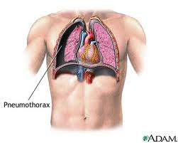
Pneumothorax (Collapsed lung)

Pneumothorax is the obtainment of air in the pleural cavity.
tension pneumothorax, spontaneous pneumothorax, spontaneous, pneumothorax symptoms, pneumothorax treatment , open pneumothorax, pneumothorax symptoms, atelectasis, symptoms of pneumothorax, pneumothorax definition, right pneumothorax, pneumothorax causes, pneumothorax, what is pneumothorax, Hemopneumothrax

Pathophysiology and Etiology
  1. Spontaneous pneumothorax due largely located superficial bullae rupture, and preceded by an increase in intra-pulmonary pressure, among others: cough hard or after blowing musical instruments, sneezing, straining, and others. The entry of air into the pleural cavity, through a tear in the visceral pleura. Bula congenital, predisposing especially in young males. Bula may also arise due to pulmonary tuberculosis, pneumoconiosis and bronchial obstruction.
  2. Traumatic pneumothorax can be caused by a "penetrating and non penetrating injury," either with or without rib fractures.
  3. "Surgical trauma" and "iatrogenic damage". Surgery can cause this type of pneumothorax.
  4. "Artificial pneumothorax" necessary for the treatment of hemoptysis in pulmonary tuberculosis as well as diagnostic measures for lung tumors.
  5. "Tension pneumothorax" due to "check valve" mecanism, so that air can enter but can not get out of the pleural cavity to the mediastinum due to a healthy one side driven.
  6. Open pneumothorax, occurs when the visceral pleura remains torn open so that the pressure in the pleural cavity with the outside air pressure.
  7. Closed pneumothorax, when ripping a hole closes after the incoming air is quite a lot. As a result the pressure in the pleural cavity is higher than the outside air pressure.
  8. Hemopneumothoraks, hidropneumothoraks and piopneumothoraks, when the pleural cavity contains blood, pleural fluid or pus clear.

Symtoms and Clinical Examination

  1. Sudden chest pain
  2. Sudden shortness of breath
  3. Respiratory failure and may be accompanied by cyanosis Puls.
Physical examination
  1. There is often a "Circulatory collapse" because of "Tension pneumothorax".
  2. In the percussion sound obtained hipersonor
  3. On auscultation, decreased breath sounds found on the side until the pain disappears.

Chest X-ray Pictures
1. On PA X-ray chest images visible edge of a collapsed lung in the form of a line. In pneumothorax parsialis the localization in the anterior or posterior, the perimeter of the lung may not be visible.
2. "Mediastinal shift" can be seen in the photograph Anatotomical Pathology or fluoroscopy when the patient inspiration or expiration, especially can occur in "Tension pneumothorax".

Differential Diagnosis
1. Pleurisy and pericarditis
2. Myocardial infarction and pulmonary embolism
3. Chronic bronchitis and emphysema
4. "Diaphragmatic herniae"
5. "Dissecting aneurysmae aortae".

Indication-specific treatment:
1. "Tension pneumothorax"
2. Pneumothorax accompanied by shortness of breath.
3. Bilateral pneumothorax
4. Large pneumothorax. When a line edge of a collapsed lung> 1 / 3 transverse diameter.
5. There appear to pleural fluid accumulation are aplenty
6. Pneumothorax as a complication of pulmonary diseases, such as supurativa pneumonia, tuberculosis.
7. Recurrent pneumothorax
8. Complications in the use of a ventilator.

Procedure and Management Of Pneumothorax

Complications
  1. "Tension pneumothorax" will berakhur fatal, if there is "Circulatory collapse".
  2. Respiratory failure.
  3. Hemopneumothrax
  4. Secondary infections
  5. Pleural thickening
  6. Atelectasis
  7. Recurring. 20% in simple or pneumothraks pneumothraks simplex and 50% in patients with COPD
  8. Mediastinal emphysema
  9. "Re-expansion pulmonary oedema".
Prognosis
  1. Good, if immediate relief and intensive treatment, especially regarding healthy young patients.
  2. Essentially dependent disease or "underlying disease", is very dangerous when the patient with COPD.

Reference

  1. Crofton J, Douglas A. Respiratory Disease, 3rd edition . Blackwell Scientific Publications, Singapore, 1984, pp. 541 – 546.
  2. Graham K, Crompton. Diagnosis and Management of Respiratory Diseases. Blackwell Scientific Publications. Oxford-London- Edinburg 1980, p. 150 – 163.
  3. Sol Katz. Spontaneus Pneumothorax In : Respiratory Emergency. Ed. By Moser KM, Spragg, R.G, 2nd edition. The CV Mosby Company, London, 1982, p. 176 – 193.

Tags : tension pneumothorax, spontaneous pneumothorax, spontaneous, pneumothorax symptoms, pneumothorax treatment , open pneumothorax, pneumothorax symptoms, symptoms of pneumothorax, pneumothorax definition, right pneumothorax, pneumothorax causes, pneumothorax, what is pneumothorax, Hemopneumothrax

Related Posts Plugin for WordPress, Blogger...
Ping your blog, website, or RSS feed for Free TopOfBlogs

Followers