Definition | Treatment | Management of Disease | Diagnosis | Symptoms | Etiology | Pathophysiology | Procedure

 
Showing posts with label Management Of Disease. Show all posts

Management of Cough

The best management of cough is best to specific drug delivery to the etiology. Three forms of management of cough are:

1. Without the drug delivery
Cases with a cough without the interference caused by acute illness and heal itself usually does not need medication.
2. Specific Treatment
This treatment is given to the causes of cough.
If the cause of cough is known then the treatment should be directed towards the cause. With an integrated diagnostic evaluation, in almost all patients can be a known cause of chronic cough.
Specific treatment depends on the etiology or the cough mechanism. Asthma treated with bronchodilators or corticosteroids. Post nasal drip due to sinusitis treated with antibiotics, nasal spray and antihistamine-decongestant combinations, post nasal drip due to allergies or non allergic rhinitis dealt with avoiding environments that have the precipitating factors and antihistamine-decongestant combinations.
Gastroesophageal reflux treated by elevating the head, dietary modifications, antacids and cimetidine. Cough in chronic bronchitis treated by stopping smoking. Antibiotics are given to pneumonia, sarcoidosis treated with corticosteroids and cough in congestive heart failure with digoxin and furosemide.
Specific treatment also may include surgery such as pulmonary resection in lung cancer, polypectomi, remove hair from the outer ear canal.
Cases with a cough without the interference caused by acute illness and heal itself usually does not need medication.
3. Symptomatic treatment
Given both to patients who can not determined the cause of the cough as well as to patients who cough is a nuisance, not working properly and can potentially cause complications.
Symptomatic treatment is given if:
The cause of cough is certainly not known, so that specific treatment can not be given.
Coughing is not functioning properly and its complications endanger the patient.
Drugs used for symptomatic treatment of two types namely:
a. Antitussive
Antitussive is a medication that suppress the cough reflex, used in respiratory disorders and unproductive coughs due to irritated skin.
In general, based on place of work is divided into antitussive  drug that works in the peripheral and central antitussive who works at. Working in the central antitussive divided into non-narcotic and narcotic.
b. Mucokinesis
A pathologic fluid retention in the airway is called mucostasis. Drugs that are used to handle the situation called mucokinesis.

See more : 

Management of COPD

The goal of COPD management are:
  1. Touch ups the ability of people with symptoms mengatasiu not only in the acute phase, but also the chronic phase.
  2. Improving the ability of the patient in carrying out daily activities.
  3. Reducing the rate of progression of disease if the disease can be detected early.


Management of COPD in the elderly are as follows:
  1. Etiological factors negate / precipitation, for example immediately stop smoking, avoid air pollution.
  2. Cleaning the bronchial secretions to help in various ways.
  3. Eradicate the infection with antimicrobial. In the absence of antimicrobial infections need not be given. Provision of appropriate antimicrobial should be in accordance with the germs that cause infections according to the results of sensitivity testing or empirical treatment.
  4. Overcome bronchospasm with bronchodilator drugs. The use of corticosteroids to resolve the inflammatory process (bronchospasm) is still controversial.
  5. Symptomatic treatment.
  6. Treatment of the complications that arise.
  7. Oxygen treatment, for those who need. Oxygen should be administered with a slow flow of 1-2 liters / minute.
  8. Rehabilitation actions which include:
  • Physiotherapy, mainly aims to help the expenditure of bronchial secretions.
  • Breathing exercises, to train the patient in order to make the most effective breathing.
  • Exercise with weights oalh particular sport, with the aim to restore physical fitness.
  • Vocational guidance, the work done against the patient can re-do their previous occupation.
  • Psychosocial management, primarily intended for patient self-adjustment to her illness.

 COPD (Chronic Obstructive Pulmonary Disease)

Tags : Management of COPD, COPD treatment, The goal of COPD management,

Management of Occupational asthma

To prevent the occurrence of occupational asthma is a medical examination prior to employment, use of protective equipment, monitoring air pollutants in the working environment is highly recommended. If occupational asthma has occurred, then transfer to the outside of the work environment is important. If for some reason can not be moved then it must be the prevention and monitoring of lung function decline.

Periodic evaluation of lung function in workers who already suffer from occupational asthma are needed to prevent disability. Clinical asthma will persist until a few years even though the worker has been out of work environment.


Medical treatment in patients with occupational asthma such as bronchial asthma in general:

  1. Theophylline, a bronchodilator and may suppress neutrophil chemotactic factor. Effectiveness of the two functions above depend on the serum levels of theophylline.
  2. Beta agonists, bronchodilators are the most good for the treatment of occupational asthma in comparison with cholinergic antagonists (ipratropium bromid).
  3. The combination of beta-agonists with ipratropium bromid improve lung function is better than just a beta agonist alone.
  4. Corticosteroids, from various studies is known to prevent the bronchoconstriction caused by bronchial provocation with allergens. It will also improve lung function, decrease exacerbations and airway hiperesponsivitas and will ultimately improve the quality of life.

Management Of Pleural Effusion

Management Of pleural Effution :
  • Pleural fluid aspiration performed, to reduce the unpleasant taste or "discomfort" and shortness of breath.  It is advisable to gradually aspirations.Fluids released between 500 - 1000 cc. When taking too many and can quickly lead to pulmonary edema.
  • Incorporating intrapleura chemotherapy for malignancy (eg radioactive gold or Ytrium).
  • More often performed pleurodesis in the process of malignancy or in the frequent recurrence of effusion.
  • By using 500 mg tetracycline powder is dissolved in 50 cc of physiologic saline. Patients shake it so flat, then the liquid removed after clamped for 24 hours or given jodium powder or talc.
  • Pain that occurs because the administration of drugs above can be overcome with the analgesic. If need be given pethidine 100 mg i.m.
  • Provision of steroids combined with antituberculosis can absorb the pleural effusion caused by pulmonary tuberculosis quickly and reduce fibrosis.

 Management Of Pleural Effusion,Pleural effution, Pleural effussion, treatment pleural effusion,  Pleural effusion Procedure, left pleural effusion


Tags : Pleural effutionPleural effussion, treatment pleural effusion,  Pleural effusion Procedureleft pleural effusion

Management of Chronic Bronchitis

General:
  • Education to patients and their families
  • Stop smoking and avoid air pollution
  • Prevent infection
  • Clean environment
  • Hydration moderation: drink enough water (8-10 glasses a day)
  • Proper nutrition: protein rich diet and avoid heavy meals before bedtime, milk can cause increased bronchial secretions, should be prevented.


Provision of drugs:
1. Bronchodilators:
  • Aminophylline inj. 5.6 mg / kg i.v. or oral loading dose of 3 x 100-200 mg.
  • Terbutaline 3 x 2.5 mg oral or injection 0.25 mg s.c. every 4-6 hours (1 mg / ml; = 2 ml ampoule).
  • 3 x 2 mg salbutamol orally.

2. Expectorant:

  • Water is a good expectorant
  • Glyceryl guaiacolate 4 x 100-200 mg.

3. Mucolytic :

  • Bromhexine HCL: 3 x 1 tablet by mouth.
  • N-acetyl cysteine: 3 x 200 mg orally.

4. Respiratory therapy:

  • Aerosols: Ipratropium Bromide 3 x 2 puffs
  • Oxygen: 1-2 liters / minute via nasal cannula if PaO2 <55 mm Hg.
5. Rehabilitation:
  • Physiotherapy
  • Relaxation exercises
  • Breathing exercises
  • Chest percussion and postural drainage
  • Exercise physical abilities
  • Psychosocial Rehabilitation
  • Vocational Rehabilitation.

Management Of Hemothorax

Hemothorax patient death can be caused because of the large blood loss and the occurrence of respiratory failure.
Respiratory failure due to the large amount of blood in the pleural cavity pressure of lung tissue and reduced lung tissue that does ventilation.

Then treatment hemothorax as follows:
1. Emptying of blood from the pleural cavity.
Installed "chest tube" and is connected with the WSD system, this can accelerate the lung expands.
2. Stop the bleeding.

If the installation of WSD, the blood still does not stop, then considered for thoracotomy.
3. General state of repair.
Giving oxygen 2-4 liters / minute, the length adjusted to the clinical changes, better yet, if the monitored with blood gas analysis. Try to people with normal blood gases.
Giving blood transfusion: seen from a decrease in Hb.
As a benchmark can be used the following calculation, every 250 cc of blood (from patients with Hb 15 g%) can raise ¾ g% Hb.
Given with a normal drop of about 20-30 drops / minute and maintained not to an interruption in heart function or cause interference with the heart.
4. Others.
  • Antibiotics, carried out if there is secondary infection.
Antibiotics are used adjusted to the sensitivity test and culture. If the infecting organism is unclear, while the state of serious illnesses, then the patient can be given a "broad spectrum antibiotic", for example, ampicillin at a dose of 4 x 250 mg daily.
Also considered in case of pleural thickening decortication.

Tags : Management Hemothorax, Hemothorax procedure, Thoracotomy, hemothorax treatment, haemothorax, hemothorax

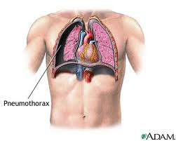
Management Of Pneumothorax

Outside the hospital.
  1. In light of spontaneous pneumothorax or pneumothorax simplex. Minimal or no complaints at all, are usually found by accident. The air in the pleural cavity will diresorbsi spontaneously. Because it does not require invasive measures.
  2. "Tension pneumothorax". Done in a sterile and carried out the stabbing in the sore area with a syringe the size of the largest. Stabbings in the space between the ribs into 2 in the front line of mid-clavicle. In young women (cosmetics) stabbings in the space between the ribs into 4 or 5 in the mid-axillary line. Then the needle tip covered with a sheet of thin rubber or thin plastic that can serve as a valve. Subsequently the patient was sent to hospital.
In the hospital.
  1. At the same place to do the installation of WSD, using trokar (troicar). It should be noted, that all actions undertaken SCARA sterile.
  2. WSD is removed, when the lung is expanding well and no complications after plastic hose clamped shut or 24 hours to prove that the pneumothorax was cured.
  3. If the patient is congested, it can be administered with high concentrations of oxygen and given to people with healthy lungs (before). In patients with COPD oxygen delivery must be careful.
  4. To treat pain may be given analgesics like-antalgin 3 x 1 tablet.
  5. In pneumothorax with severe COPD, is sometimes given strong analgesics such as pethidin 100 mg im or morphine 10 mg i.m. Physiotherapy should be given, because it could prevent sputum retention.
  6. If the lung development is rather slow, can be done with a suction pressure of 25-50 cm of water.
  7. In a recurrent pneumothorax (recurrent) do both pleural adhesions by using a material that can cause irritation or materials "scleroting agent".
  8. If there is a-Bronco-pleural fistula, it will be done eksterpasi operation.

Management of Empyema Thoracic

The principle of treatment in empyema:
1. Emptying of the pleural cavity of pus
2. Antibiotics
3. Closure of the pleural cavity
4. Causal treatment
5. Additional treatment.

1. Emptying of the pleural cavity.
a. Simple aspiration.
Performed repeatedly using a large needle hole. This method is good enough to remove most of the pus or fluid from acute empyema is still runny.
Losses such as these techniques often lead to "pocketed" empyema.
Ultrasound can be used to determine the localization of "pocketed" empyema.
b. Drainage is closed.
Installation "= closed thoracostomy tube drainage (WSD)".
Indications of this drain fitting, if the pus is very thick, pus is formed after 2 weeks and there has been piopneumotoraks.
Installation of the hose should not be too low, the diaphragm is usually raised because of empyema. Select a hose that is large enough.
If 3-4 weeks of no progress should be pursued by other means, such as in chronic empyema.
c. Installation of "open-drainage":
This action is done on a chronic empyema by cutting a piece of rib to create a "window". This method is chosen when dekortikasi not possible and should be done in a completely sterile condition.


2. Antibiotics.
Given the major cause of death due to sepsis, then antibiotics play an important role.
Antibiotics should be given immediately once the diagnosis is established and the diagnosis should adcquate. The selection of antibiotics based on results of Gram staining of pus smear.
Subsequent treatment depends on the culture results and sensitivity tests.
When germs that cause is unclear, can be used high doses of benzyl penicillin.
3. Closure of the pleural cavity.
When chronic empyema fails to show a response to the drainage hose, then do dekortikasi or torakoplasti.
If not handled properly will add to the long hospitalization.
4. Causal treatment.
Depending on the cause eg, amubiasis, tuberculosis, aktinomikosis.
Treated with specific drugs for each disease.
5. Additional treatment and physiotherapy.
Aiming to improve the general situation

Tags = Empyema treatment, Antibiotic for EmpyemaManagement Of empyema thoracic, management for empyema thoracic, empyema thoracic management, empyema thoracic treatment, treatment of empyema thoracic, empyema thoracic medical management, Management Of empyema thoracis, management for empyema thoracis, empyema thoracis management, empyema thoracis treatment, treatment of empyema thoracis, empyema thoracis medical management

Management of Bronchiectasis

A. Conservative:
1. Control of acute and chronic infection, mucus secretion, airway obstruction and complications, such as: coughing up blood, respiratory failure and cor pulmonale, in order to prolong life, improve quality of life and prevent disease progression.
2. Antibiotics if there is infection.
3. Chest physiotherapy and postural drainage with forced expiratory technique for removing secretions.
4. Aerosols with physiologic saline or beta agonists prior to chest physiotherapy may facilitate the release of sputum / secretions.
5. Bronchodilators to improve airflow, helping mukosilia clearance and physiotherapy improve outcomes.
6. Corticosteroids when there is severe bronchospasm (CPOD or Asthma Bronchiale).
bronchiectasis pneumonia, what is bronchiectasis, bronchiectasis copd, bronchiectasis symptoms, bronchiectasis treatment, bronchiectasis definition, cystic bronchiectasis, Management bronchiectasis, Procedure of bronchiectasis
B. Surgery:
Indications of surgery:

Tags : bronchiectasis pneumonia, what is bronchiectasis, bronchiectasis copd, bronchiectasis symptoms, bronchiectasis treatment, bronchiectasis definition, cystic bronchiectasis, Management bronchiectasis, Procedure of bronchiectasis

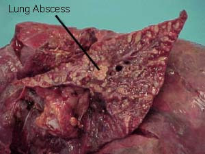
Management of Lung abscess

1. General management:
Improving the general condition of patients with high-calorie high-protein diet and drink plenty of fluids.
a. Antibiotics.
  • Procaine penicillin G given 1.2 million units im every 12 hours + chloramphenicol 500 mg every 6 hours for 10-15 days. or
  • Procaine penicillin G 1.2 million units i.m. every 12 hours + Metronidazole 500 mg every 6 hours for 10-15 days. or
  • Clindamycin 600 mg every 8 hours for 10-15 days.

b. Postural drainage and physiotherapy.
The position of the body are arranged so that pus can come out by itself (due to gravity) or with the help of the physiotherapist.

2. Special Treatment:
a. Bronchoscopy
If pus is difficult exit, it is necessary to bronchoscopy to clear the airway and sucking pussy.
b. Surgery
When chemotherapy failed. A chronic abscess, cavity remains and sputum production remained there while the clinical symptoms are still present after adequate therapy for 6 weeks or the rest of extensive scar tissue that can interfere with lung physiology. This is all an indication of surgery.

Tags : Management Of Lung abscess, management for Lung abscess, Lung abscess management, community acquired Lung abscess, Lung abscess treatment, treatment of Lung abscess, Lung abscess medical management

Management of Bacterial Pneumonia

Indications of hospitalization is:
1. Patients with basic diseases
2. Patients with complications.

General management.
1. Correction of underlying abnormalities (underlying disease)
2. Bedrest
3. Symptomatic medications, administered only if necessary, such as:
  • 3 x 500 mg Paracetamol (in hyperpyrexia)
  • Morphine 10 mg s.c. (If there is pain severe).

4. Maintain fluid and electrolyte balance with the help of intravenous fluids, 5% dextrose, normal saline or Ringer's lactate.

5. Selection of anti-infective drugs.
Selection of antimicrobial drugs, should be based on sensitivity tests and sensitivity, but due to time and facilities greatly affect the success of this test, then giving more medicine based on empirical, as below:

Special Treatment:
1. Pneumococcal pneumonia,
Basic disease: elderly, Chronic Obstructive Pulmonary Disease (COPD), CHD, diabetes mellitus, alcoholism, post-influenza.
Clinical features: sudden illness, high fever, chills, pleuritic pain, cough productive with phlegm such as iron rust, herpes labialis.
Physical diagnostics: signs of consolidation.
Chest X photo: visible presence consolidated picture.
Laboratory:
- Blood banks: lekositosis 15,000 - 40.000/ml.
- Sputum: gram positive cocci which are lancet-shaped.
Treatment:
a. Antibiotics options:
Penicillin G from 0.6 to 1.2 million units i.m. every 12 hours, for 50-10 days.
b. Antibiotics alternatives:
Erythromycin 500 mg orally every 8 hours for 5-10 days or Tetracycline 500 mg orally every 8 hours for 5-10 days.

2. Staphylococcal pneumonia.
Basic disease: post-influenza, diabetes mellitus, malignancy, advanced age, use of drugs.
Clinical features: sudden onset of illness accompanied by high fever, chills, pleuritic pain and shortness of breath accompanied by cough productive with purulent sputum (with blood).
Physical diagnostics: look for signs of consolidation in one lobe or
several segments, and quickly arise necrosis, abscess and
pneumatokel.
Chest X photo: a picture of consolidation in one lobe or the
some segments.
Laboratory:
- Blood banks: leucocytes 15,000 - 35.000/ml
- Sputum: gram-positive cocci found that gather.
Treatment:
a. Antibiotic choice: cloxacillin 250-500 mg iv given every 6 hours for 5-10 days.
b. Alternative antibiotics: Cephalexin / Sefazolin 0.5 to 1 gram, 3 times a day, for 50-10 days.

3. Pneumonia klebsiela.
Basic diseaseChronic Obstructive Pulmonary Disease (COPD), alcoholics, elderly, diabetes mellitus
Clinical symptoms: sudden illness, high fever, chills, pleuritic pain, shortness of breath, productive cough with phlegm like jelly (currant jelly)
Physical diagnostics: according to the signs of consolidation, chest looks
more convex.
Chest X photo: picture looks consolidation with an enlarged volume
so that the fissure becomes convex (bulging).
Laboratory:
- Blood banks: leucocytes 15,000 - 40.000/ml.
- Sputum: gram-negative bacilli appear in the form of fat.
Treatment:
a. Antibiotic choice: Ampicillin 1000 mg i.v. given every 6 hours, plus Gentamicin 60-80 mg every 8 hours, for 50-10 days.
b. Alternative antibiotics: Cefotaxime 1 g i.v. every 6 hours for 5-10 days.

Tags : management of pneumonia, management for pneumonia, pneumonia management, community acquired pneumonia, pneumonia treatment, treatment of pneumonia, pneumonia medical management

Management Bronchial Asthma

Procedure Of Bronchial Asthma
Non-pharmacotherapy
  • Provision of O2
  • Fluid
  • Postural Drainage
  • Avoiding exposure to allergens
  • Guidance on patients and families about asthma, causes, and how to overcome them.
  • Avoid the trigger factors (diet, medication, living habits, allergens)
  • Immunotherapy / desentisisasi
  • Physiotherapy breath, vibration and / or thoracic percussion, an efficient cough.

Management of asthma , management of asthma bronchial , procedure of asthma,  asthma bronchial  Procedure
Pharmacotherapy:
1. Bronchodilators:
- Adrenaline; solution of adrenaline 1: 1000 subcutaneous
0.3 cc waiting for 15 minutes, if it has not subsided again given
0.3 cc if it has not abated, can be repeated once again 15 minutes later
0.3 cc.
For children can be given smaller doses: 0.1 to 0.2 cc.
Caution in elderly patients (coronair heart disease), hypertension, hyperthyroidism.
-  Beta 2 agonists (oral, injection, inhalation / MDI, nebulizer)
Orsiprenalin (alupent) 3 x 20 mg orally; subcutan 3 x 0.25 mg; 3 x 3 spray inhalation.
Bricasma subcutan 3 x 0.5 cc.
Heksoprenalin (Ipradol) 3 x 0.50 mg orally.
Salbutamol 2 mg orally 3 times.
- Methylxanthine (oral, suppository, injection):
a. Aminophyllin, given intravenously slowly 5-10 minutes, give 5-10 cc.
b. Aminophyllin can be granted if after 2 hours by administration of adrenaline did not give results.
c. Aminophyllin drip / iv 3 x 250 mg, drip: loading dose 5 mg / kg, followed by drops of 0.9 mg / kg / hour, or 20 mg / kg BB/24 hours.
d. Oral Aminophyllin 3 x 120-150 mg.
- Anticholinergics
a. 3 x 0.25 mg atropine subcutaneously
b. Ipatroprium bromid 3 x 2 puffs metered dose aerosol.
2. Anti-inflammatory:
- Corticosteroids (oral, injection, aerosol form of metered-dose inhaler / MDI):
- Intravenous dexamethasone 3 x 1 ampoule
3 x oral prednisone 50-10 mg, once achieved dose effects lowered slowly
- cromolyn (sodium cromoglycate), nedocromil and others.
3. Antihistamines:
- CTM 3 x 2-4 mg.

4. Antibiotics are given if there is an infection:
-  4 x 250 mg Amoxicillin
-  4 x 250 mg Tetracycline
-  Cotrimoksazole 2 x 2 tablets
-  Erythromycin  3 x 250 mg

5. Expectorant - mucolytic:
- Ordinary drinking water (diluent secretions)
- Glyceril guaiacolat (expectorans)
- Potassium Jodide (expectorans)
- N-Acetyl-cysteine ​​(secretolytic)
6. Immunotherapy:
Accomplished by repeated subcutaneous injection, allergen extracts with increasing concentrations, in patients with allergic asthma due to high levels of IgE.

How to use inhalers (MDI) is right:
  1. MDI opened the lid, shake 3-4 times
  2. Place in front of the mouth is open wide (4 cm) or a "spacer" that is inserted in the mouth.
  3. Perform up to a maximum expiratory then push the MDI, while spraying the medicine, do inspire slowly for 5 seconds or more.
  4. When it reaches a maximum inspiration, hold breath for 10 seconds.
  5. Then breath usual 3-5 minutes. Further repeated measures a. when needed.
  6. We recommend that you rinse after using the inhaler.

Reference :

  1. Brashear RE. Chronic Obstructive Lung Disease. Clinical Treatment and Management. Mosby Co, St Louis, 1982, pp. 4 – 17.
  2. Baum GL. Textbook of Pulmonary disease. 3 rd ed. Little Brown Co. Boston, 1983. pp. 405 – 407.
  3. Harrison’s Principles of Internal Medicine. 10 th ed. McGraw Hill, Tokyo, pp. 1512 – 1519.
  4. Kay AB. Asthma : Clinical Pharmacology and therapeutic Progress. 1 st ed. Blackwell Scientific Publ. London, 1986, pp. 28 – 282.
  5. Sluiter HJ. Leerboek Longziekten. Van Gorcum, Assen, 1985, pp. 191 – 224.
Special Treatment of Bronchial Asthma

Related Posts Plugin for WordPress, Blogger...
Ping your blog, website, or RSS feed for Free TopOfBlogs

Followers