Definition | Treatment | Management of Disease | Diagnosis | Symptoms | Etiology | Pathophysiology | Procedure


Management Of Hemothorax

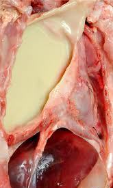
Hemothorax patient death can be caused because of the large blood loss and the occurrence of respiratory failure.
Respiratory failure due to the large amount of blood in the pleural cavity pressure of lung tissue and reduced lung tissue that does ventilation.

Then treatment hemothorax as follows:
1. Emptying of blood from the pleural cavity.
Installed "chest tube" and is connected with the WSD system, this can accelerate the lung expands.
2. Stop the bleeding.

If the installation of WSD, the blood still does not stop, then considered for thoracotomy.
3. General state of repair.
Giving oxygen 2-4 liters / minute, the length adjusted to the clinical changes, better yet, if the monitored with blood gas analysis. Try to people with normal blood gases.
Giving blood transfusion: seen from a decrease in Hb.
As a benchmark can be used the following calculation, every 250 cc of blood (from patients with Hb 15 g%) can raise ¾ g% Hb.
Given with a normal drop of about 20-30 drops / minute and maintained not to an interruption in heart function or cause interference with the heart.
4. Others.
  • Antibiotics, carried out if there is secondary infection.
Antibiotics are used adjusted to the sensitivity test and culture. If the infecting organism is unclear, while the state of serious illnesses, then the patient can be given a "broad spectrum antibiotic", for example, ampicillin at a dose of 4 x 250 mg daily.
Also considered in case of pleural thickening decortication.

Tags : Management Hemothorax, Hemothorax procedure, Thoracotomy, hemothorax treatment, haemothorax, hemothorax

Hemothorax

Hemotorax is the presence of blood in the pleural cavity.

Pathophysiology and Etiology
  1. Sharp trauma or blunt trauma. This can occur when blunt trauma can cause fractures of the ribs, resulting in a torn intercostal blood vessels and also cause a tear in the lung tissue.
  2. A torn aortic aneurysm
  3. Complications due to drug administration on pulmonary infarction antikoagulansia
  4. In patients with abnormalities of "hemorrhagic diathese".
  5. Complications in thoracic surgery.
Clinical Symptoms


Symptoms and complaints hemothorax depending on the weight and severity of trauma. Patients may complain of shortness of breath, chest pain, until the shock and anemia.

Diagnosis
1. Anamnesa : A history of trauma to the chest, or after surgery.
2. Physical examination
  • Found such signs in the pleural effusion.
  • At the hospital hemitoraks reduced movement.
  • Hemithorax percussion on the sick and faint sounds on auscultation, breath sounds audible reduced or disappear altogether.
3. Chest X-ray photos : Radiological picture as in pleural effusion.
4. Laboratory.
After aspiration of the experiment, the liquid is carried out the examination as follows:
  • If the number of erythrocyte 5000-6000 / mm 3 --> "a Rosy tint".  If the number of red blood cells> 10,000 / mm 3 --> "serosanguinous". It is found in pleural effusion hemoragic.
  • If the fluid coming from complications hemoragis torasentesis, the liquid is centrifuged and the supernatant became clear. In contrast to the supernatant hemothorax still red.
  • It is said hemothorax tilapia Hb blood from the pleural cavity> 1 g / dl or if the hemoglobin derived from blood hemothorax half the price of capillary blood Hb.
Management Of Hemothorax

Complications
  1. Loss of blood.
  2. Respiratory failure.
Tags :definition hemothorax, hemothorax pleural effusion, hemothorax symptoms, tension hemothorax, hemothorax treatment, hemothorax chest tube, what is hemothorax, Management Hemothorax, Complications Hemothorax, hemothorax therapy, hemothorax procedure, Hemotorax, Haemothorax , Etiology Hemothorax, Pathophysiology Hemothorax

Management Of Pneumothorax

Outside the hospital.
  1. In light of spontaneous pneumothorax or pneumothorax simplex. Minimal or no complaints at all, are usually found by accident. The air in the pleural cavity will diresorbsi spontaneously. Because it does not require invasive measures.
  2. "Tension pneumothorax". Done in a sterile and carried out the stabbing in the sore area with a syringe the size of the largest. Stabbings in the space between the ribs into 2 in the front line of mid-clavicle. In young women (cosmetics) stabbings in the space between the ribs into 4 or 5 in the mid-axillary line. Then the needle tip covered with a sheet of thin rubber or thin plastic that can serve as a valve. Subsequently the patient was sent to hospital.
In the hospital.
  1. At the same place to do the installation of WSD, using trokar (troicar). It should be noted, that all actions undertaken SCARA sterile.
  2. WSD is removed, when the lung is expanding well and no complications after plastic hose clamped shut or 24 hours to prove that the pneumothorax was cured.
  3. If the patient is congested, it can be administered with high concentrations of oxygen and given to people with healthy lungs (before). In patients with COPD oxygen delivery must be careful.
  4. To treat pain may be given analgesics like-antalgin 3 x 1 tablet.
  5. In pneumothorax with severe COPD, is sometimes given strong analgesics such as pethidin 100 mg im or morphine 10 mg i.m. Physiotherapy should be given, because it could prevent sputum retention.
  6. If the lung development is rather slow, can be done with a suction pressure of 25-50 cm of water.
  7. In a recurrent pneumothorax (recurrent) do both pleural adhesions by using a material that can cause irritation or materials "scleroting agent".
  8. If there is a-Bronco-pleural fistula, it will be done eksterpasi operation.

Management of Empyema Thoracic

The principle of treatment in empyema:
1. Emptying of the pleural cavity of pus
2. Antibiotics
3. Closure of the pleural cavity
4. Causal treatment
5. Additional treatment.

1. Emptying of the pleural cavity.
a. Simple aspiration.
Performed repeatedly using a large needle hole. This method is good enough to remove most of the pus or fluid from acute empyema is still runny.
Losses such as these techniques often lead to "pocketed" empyema.
Ultrasound can be used to determine the localization of "pocketed" empyema.
b. Drainage is closed.
Installation "= closed thoracostomy tube drainage (WSD)".
Indications of this drain fitting, if the pus is very thick, pus is formed after 2 weeks and there has been piopneumotoraks.
Installation of the hose should not be too low, the diaphragm is usually raised because of empyema. Select a hose that is large enough.
If 3-4 weeks of no progress should be pursued by other means, such as in chronic empyema.
c. Installation of "open-drainage":
This action is done on a chronic empyema by cutting a piece of rib to create a "window". This method is chosen when dekortikasi not possible and should be done in a completely sterile condition.


2. Antibiotics.
Given the major cause of death due to sepsis, then antibiotics play an important role.
Antibiotics should be given immediately once the diagnosis is established and the diagnosis should adcquate. The selection of antibiotics based on results of Gram staining of pus smear.
Subsequent treatment depends on the culture results and sensitivity tests.
When germs that cause is unclear, can be used high doses of benzyl penicillin.
3. Closure of the pleural cavity.
When chronic empyema fails to show a response to the drainage hose, then do dekortikasi or torakoplasti.
If not handled properly will add to the long hospitalization.
4. Causal treatment.
Depending on the cause eg, amubiasis, tuberculosis, aktinomikosis.
Treated with specific drugs for each disease.
5. Additional treatment and physiotherapy.
Aiming to improve the general situation

Tags = Empyema treatment, Antibiotic for EmpyemaManagement Of empyema thoracic, management for empyema thoracic, empyema thoracic management, empyema thoracic treatment, treatment of empyema thoracic, empyema thoracic medical management, Management Of empyema thoracis, management for empyema thoracis, empyema thoracis management, empyema thoracis treatment, treatment of empyema thoracis, empyema thoracis medical management

Empyema Thoracic

Empyema Thoracic is the presence of pus in the cavity / pleural cavity.

Etiology of Empyema Thorasic
I. Derived from the lungs:
  1. Pneumonia
  2. Lung abscess
  3. The existence of bronchopleural fistula
  4. Bronchiectasis
  5. Pulmonary Tuberculosis
  6. Lung fungus.

II. From the extrapulmonary infection:
  1. Trauma of the brain
  2. Brain surgery
  3. Torasentesis
  4. Subfrenik abscess
  5. Due to amoebic liver abscess.

Bacteriology
  1. Staphylococcal piogenes, at all ages, often in children.
  2. Piogenes streptococcus.
  3. Gram-negative bacteria (Pseudomonas aeruginosa,, Klebsiela, Bakteroides, E. coli, Proteus mirabilis)
  4. Anaerobic bacteria.

Pathophysiology Of Empyema Thorasic
Due to pyogenic bacteria invasion into the pleura arising acute inflammation, followed by the formation of serous exudates. With the number of PMN cells either living or dead, as well as increased levels of protein, the fluid becomes cloudy and thick. Fouled fibrin will form pockets of pus to localize it.

Clinical Symptoms
Clinical Course
Divided into 2 stages, namely: acute and chronic.
1. Acute empyema:
Symptoms are similar to pneumonia, high fever, pleuritic pain when the stage is left in a few weeks there will be toxemia, anemia and clubbing. If pus is not issued soon will arise fistula bronkopleura and "empyema necessitatis".
2. Chronic empyema:
Strict boundary between acute and chronic hard set, called chronic empyema when running more than 3 months.
Patients complain of a weak body, health, the patient looked back, pale and no clubbing.

Diagnosis
Physical examination.
Found signs of fluid, accompanied by the movement of sick hemitoraks reduced. There was a faint sound on percussion. On auscultation, breath sounds decreased until it disappears in the hemitoraks sick.

Chest X-ray photo.
On chest PA X-ray and lateral images obtained an image of "opacity" that indicate the presence of fluid with or without pulmonary abnormalities.
In the event of fibrotoraks, trachea and mediastinum attracted to the side of the hospital and also looked pleural thickening.

Sure Diagnosis
Pleural aspiration would indicate the presence of pus in the pleural cavity.
Furthermore, the pus is used as material for examination: cytology, bacteriology fungi, amoeba, done culture (culture) and sensitivity to antibiotics.

Management of Empyema Thoracic


Complications of Empyema Thoracic
Complication that often arises is: bronchopleural fistula.
Other complications that may occur are: shock, sepsis, congestive heart failure and otitis media.


Tags : empyema thoracis, Definition Empyema, Empyema thoracic, Empyema symtomps, Empyema treatment, What is Empyema, Management Empyama, empyema treatment

Related Posts Plugin for WordPress, Blogger...
Ping your blog, website, or RSS feed for Free TopOfBlogs

Followers