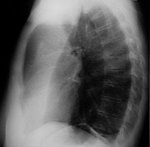
A right upper lobe collapse would appear as an opaque area at the top, with a limit that is strictly concave beneath the clavicle near the fissures caused by the raised horizontalis.
The left upper lobe usually includes Lingula when deflated, and the resulting image is less firm without the firm lower limit. But in the lateral projection would seem a tongue-shaped shadow with a peak near the diaphragm; anteriorly, it may be up to the sternum, or may be separated by a translucent area of the lungs caused by a slip right next to them and the sternum posterior shadow It has a clear boundary with concave boundary caused by large fissures are pushed to the front.
A middle lobe will cause a shadow that is not firmly on the anterior projection, but may blur the line than the right heart, in the lateral projection it will appear as a ribbon-shaped shadow extending from the hilum to the angle of the sterno-diafragmatikus. Strict upper limits established by the nearest horizontalis fissures, whereas the concave rear boundary by a major fissure is pushed forward.
Lower lobe is deflated causing a triangular-shaped shadow, with the lateral border of the firm that ran downward and outward from the hilum to the diaphragm. Therefore it is usually located behind the heart shadow, he can only be seen when the radiograph is good. On the lateral projection image may be blurred at all, but its presence usually gives three images; thoracic vertebrae at the bottom will look more gray than black than the vertebrae next to the middle; the posterior than the shadow of the left diaphragm will not be seen; and finally, vertebrae in the back area below the heart shadow will be less black than the translucent area behind the sternum.
The symptoms of the other characteristics are the consequence rather than vascular shadows have become blurred in the general opacity than lobes that do not contain air, while the shadows of blood vessels in the other lobe is more dispersed because it fills a larger volume. Hilar blood vessels on the affected side will show a disease and not a lateral convexity concafitas as in the normal state at the place where the group rather than the upper lobe artery basal met in addition, the hilum will be smaller than on the other side, while blood vessels of the lungs will be more dispersed, so per unit area will look much less than on the other side (normal). Only there will be little or no relative translucency, because of capillary flow increases, Only there will be little or no relative translucency, because of capillary flow increases, whereas the tracheal pressure or elevation of the diaphragm and heart are usually a little switch only slightly in the direction of the deflated lobe of the collapse than the lower lobe, or more often as not at all on the upper lobe collapse rather than
Comments :
0 comment to “Radiological Abnormalities of Atelectasis”
Post a Comment